
Cialis ist bekannt für seine lange Wirkdauer von bis zu 36 Stunden. Dadurch unterscheidet es sich deutlich von Viagra. Viele Schweizer vergleichen daher Preise und schauen nach Angeboten unter dem Begriff cialis generika schweiz, da Generika erschwinglicher sind.
Bio-nica.info
Genome Transplantation in Bacteria: Changing One
Species to Another
DOI: 10.1126/science.1144622
The following resources related to this article are available online at
www.sciencemag.org (this information is current as of August 3, 2007 ):
including high-resolution figures, can be found in the online
Updated information and services,
version of this article at:
can be found at:
Supporting Online Material
A list of selected additional articles on the Science Web sites
related to this article can be
found at:
, 14 of which can be accessed for free:
cites 22 articles
on August 3, 2007
This article appears in the following
Information about obtaining
of this article or about obtaining
permission to reproduce
this article in whole or in part can be found at:
Science (print ISSN 0036-8075; online ISSN 1095-9203) is published weekly, except the last week in December, by theAmerican Association for the Advancement of Science, 1200 New York Avenue NW, Washington, DC 20005. Copyrightc 2007 by the American Association for the Advancement of Science; all rights reserved. The title SCIENCE is a registered trademark of AAAS.
from incompatibility between the two genomes(6). Transplantation of nuclei as intact organelles
Genome Transplantation in Bacteria:
into enucleated eggs is a well-established proce-dure in vertebrates (7–9). Our choice of the term
"genome transplantation" comes from the sim-
Changing One Species to Another
ilarity to eukaryotic nuclear transplantation inwhich one genome is cleanly replaced byanother.
Carole Lartigue, John I. Glass,* Nina Alperovich, Rembert Pieper, Prashanth P. Parmar,
Genome transplantation is a requirement for
Clyde A. Hutchison III, Hamilton O. Smith, J. Craig Venter
the establishment of the new field of syntheticgenomics. It may facilitate construction of useful
As a step toward propagation of synthetic genomes, we completely replaced the genome of a bacterial
microorganisms with the potential to solve
cell with one from another species by transplanting a whole genome as naked DNA. Intact genomic
pressing societal problems in energy production,
DNA from Mycoplasma mycoides large colony (LC), virtually free of protein, was transplanted into
environmental stewardship, and medicine.
Mycoplasma capricolum cells by polyethylene glycol–mediated transformation. Cells selected for
Chemically synthesized chromosomes must
tetracycline resistance, carried by the M. mycoides LC chromosome, contain the complete donor
eventually be transplanted into a cellular milieu
genome and are free of detectable recipient genomic sequences. These cells that result from
where the encoded instructions can be expressed.
genome transplantation are phenotypically identical to the M. mycoides LC donor strain as judged
We have long been interested in defining a
by several criteria.
minimal genome that is just sufficient for cellularlife (10, 11) and have advocated the approach of
IthasbeenknowneversinceOswaldAvery's feature of transplantation is that the recipient chemicallysynthesizingagenomeasameansfor
pioneering experiments with pneumococcal
genome is entirely replaced by the donor ge-
testing hypotheses concerning the minimal set of
transformation more than six decades ago,
nome. There is no recombination between the
genes. The societal and ethical implications of
that some bacteria can take up naked DNA (1).
incoming and outgoing chromosomes. The result
this work have been explored (12, 13).
This DNA is generally degraded or recombined
is a clean change of one bacterial species into
Fabricating a synthetic cell by this approach
on August 3, 2007
into the recipient chromosomes to form genetic
requires the introduction of the synthetic genome
recombinants. DNA molecules several hundred
Work that is related to the process we describe
into a receptive cytoplasm. We chose myco-
kilobase pairs (kb) in size can sometimes be
in this paper has been carried out or proposed for
plasmas, members of the class Mollicutes, for
taken up. In recent studies with competent
various species. Itaya et al. transferred almost an
building a synthetic cell. This choice was based
Bacillus subtilis cells, Akamatsu and colleagues
entire Synechocystis PCC6803 genome into the
on a number of characteristics specific to this
(2, 3) demonstrated cotransformation of genetic
chromosome of a recipient B. subtilis cell using
bacterial taxon. The essential features of myco-
markers spread over more than 30% of the 4.2-
the natural transformation mechanism. The re-
plasmas are small genomes, use of UGA to en-
megabase pair (Mb) genome using nucleoid
sulting chimeric chromosome had the phenotype
code tryptophan (rather than a stop codon), and
DNA isolated from gently lysed B. subtilis proto-
of the B. subtilis recipient cell. Most of the
the total lack of a cell wall. A small genome is
plasts. Artificial transformation methods that em-
Synechocystis genes were silent (5). A schema
easier to synthesize and less likely to break
ploy electroporation or chemically competent
for inserting an entire Haemophilus influenzae
during handling. The altered genetic code fa-
cells are now widely used to clone recombinant
genome as overlapping BACs into an Escherich-
cilitates cloning in E. coli because it curtails the
plasmids. Generally, the recombinant plasmids
ia coli recipient has also been proposed; however,
expression of mycoplasma proteins. The absence
are only a few kilobase pairs in size, but bacterial
those authors have pointed out difficulties arising
of a cell wall makes the exterior surfaces of these
artificial chromosomes (BACs) greater than 300kb have been reported (4). Recombinant plas-
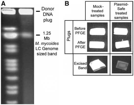
Fig. 1. Demonstration
mids coexist with host-cell chromosomes and rep-
that the DNA in the
licate independently. Two other natural genetic
blocks was intact and
transfer mechanisms are known in bacteria.
circular, whereas the
These are transduction and conjugation. Trans-
DNA in the band that
duction occurs when viral particles pick up chro-
migrated into the gel
mosomal DNA from donor bacteria and transfer
was linear. (A) A pulsed-
it to recipient cells by infection. Conjugation in-
field gel loaded with a
volves an intricate mechanism in which donor
plug containing M.
and recipient cells come in contact and DNA is
mycoides LC DNA. The
actively passed from the donor into the recipient.
1× TAE buffer gel wasseparated by electropho-
Neither of these mechanisms involves a naked
resis for 20 hours and
DNA intermediate.
then stained with SYBR
In this paper, we report a process with a dif-
gold. The marker lane
ferent outcome, which we call "genome trans-
contains Bio-Rad Saccha-
plantation." In this process, a whole bacterial
romyces cerevisiae ge-
genome from one species is transformed into
nomic DNA size markers.
another bacterial species, which results in new
Note the large amount of
cells that have the genotype and phenotype of the
DNA remaining in the
input genome. The important distinguishing
plug. (B) The plugs areshown either before PFGE or after PFGE, and the genome sized band produced after PFGE, and either with
The J. Craig Venter Institute, Rockville, MD 20850, USA.
or without treatment with the Plasmid-Safe DNase. The nuclease enzyme digests linear DNA, but has no
*To whom correspondence should be addressed. E-mail:
effect on circular duplex DNA. These data indicate the band of DNA that migrated into the gel was
exonuclease-sensitive and, therefore, linear.
3 AUGUST 2007 VOL 317 SCIENCE www.sciencemag.org
bacteria similar to the plasma membranes of
tunistic pathogens of goats, but can be grown in
At the outset, we explored a number of meth-
eukaryotic cells and may simplify our task of
the laboratory under Biosafety Level 2 condi-
ods for genome transplantation. The process had
installing a genome into a recipient cell by allow-
tions. In preparation for our experiments, it was
three key phases: isolation of intact donor ge-
ing us to use established methods for insertion of
necessary to sequence both genomes and com-
nomes from M. mycoides LC, preparation of
large DNA molecules into eukaryotic cells.
pare them to determine the degree of relatedness.
recipient M. capricolum cells, and installation of
We elected to develop our genome transplan-
We found that 76.4% of the 1,083,241-bp draft
the isolated genome into the recipient cells. We
tation methods using two fast-growing myco-
sequence of the M. mycoides LC genome (14)
chose our donor and recipient cells for genome
plasma species, Mycoplasma mycoides subspecies
could be mapped to the 1,010,023-bp M.
transplantation on the basis of our observation
mycoides, Large Colony strain GM12, and
capricolum genome (15), and this content
that plasmids containing a M. mycoides LC
Mycoplasma capricolum subspecies capricolum,
matched on average at 91.5% nucleotide identity.
origin of replication complex (ORC) can be
strain California kid, as donor and recipient
The remaining 24% of the M. mycoides LC
established in M. capricolum, whereas plasmids
cells, respectively. They divide every 80 and 100 min,
genome contains a large number of insertion
with an M. capricolum ORC cannot be es-
respectively. These organisms are both oppor-
sequences not found in M. capricolum.
tablished in M. mycoides LC (16).
Donor Genomic DNA Preparation
F i g . 2 . SDS –poly-
DNase I after Markers
Manipulation of whole chromosomes in solu-
acrylamide gel electropho-
tion exposes the DNA to shear forces that can
Intact Cells
resis (SDS-PAGE) analysis
Before or
Proteinase K
Proteinase K
cause breakage. Thus, it was important to mini-
Plug Only
of isolated M. mycoides LC
mize genome manipulation during the detergent
DNA in agarose blocks
and proteolytic enzyme treatments by suspend-
shows that there were no
ing the cells in agarose blocks. Intact chromo-
detectable proteins asso-
somes were immobilized in the resulting cavern
ciated with the DNA. The
in the agarose that originally held the cell. Di-
gels were silver-stained.
gested protein components, lipids, RNAs, and
(Left) The three lanes
sheared genomic DNAs could then be removed
labeled "Intact cells" were
by dialysis or electrophoresis from the immobi-
three dilutions of M. mycoides
on August 3, 2007
lized intact genomic DNA.
LC cells that were boiled in
Whole, intact genomic DNA isolation was
SDS and loaded onto the
performed using a CHEF Mammalian Genomic
gel. (Middle) Agarose
DNA Plug Kit from Bio-Rad. Briefly, we grew
blocks with the M. mycoides
M. mycoides LC cells containing tetracycline-
LC DNA that were boiled inSDS and loaded on the
resistance (tetM) and b-galactosidase genes
protein gel either before (B) or after (A) PFGE. (Right) To determine whether the material at the top
(lacZ) (17) at 37°C to moderate density in SP4
of the gel was protein or DNA, we treated the blocks, before and after PFGE, with DNase I. One of the
medium (18), supplemented with 10 mg/ml of
markers was DNase I.
tetracycline and, in some experiments, 10 mg/mlof streptomycin. Fifty to 100 ml of cultured cells
was reduced to a pellet by centrifugation at
Table 1. Results of a series of transplantation experiments.
4575g for 15 min at 10°C. We resuspended cellsin 20 ml of 10 mM Tris (pH 6.5) plus 0.5 M
Number of colonies
sucrose; spun as before; and resuspended again in
Negative controls
Total M. capricolum
1 ml ( 1 to 5 × 10⁹ cells/ml). We incubated the
cell suspension for 15 min at 50°C, then mixed it
with an equal volume of 2% low-melting-point
(LMP) agarose in 1× TAE buffer [40 mM Tris-
acetate and 1 mM EDTA]. After 5 min at 50°C,
the mixture of cells and LMP agarose (2 ml) was
distributed in 100-ml aliquots into plug molds.
The 20 plugs solidified at 4°C. Embedded my-
coplasma cells were lysed and proteins were
digested at 50°C for 24 hours by addition of 6 ml
of proteinase K reaction buffer [100 mM EDTA
(pH 8.0), 0.2% sodium deoxycholate, and 1%
sodium lauryl sarcosine] with 240 ml of protein-
ase K (>600 U/ml). The 20 plugs were then
washed four times at room temperature for 1 hour
in 20 ml of 1× Tris-EDTA buffer [Tris-HCl (20
mM) and EDTA (50 mM), (pH 8.0)] with
agitation and stored in 10 ml of Tris-EDTA
buffer at 4°C.
We wanted to confirm that our gentle prepa-
ration of the genomic DNA yielded intact circular
molecules. We subjected some agarose plugs to
*We attribute these two colonies to laboratory error, and we never saw any colonies on the no-donor-DNA control plates in any
pulsed-field gel electrophoresis (PFGE) in a 1%
later experiments.
†After this experiment, we did six experiments not listed here that produced no transplant clones. ‡We
LMP gel in TAE, with contour-clamped homo-
attribute the higher genome transplantation efficiency in these experiments to the inclusion of streptomycin in the SP4 mediumused to grow the M. mycoides LC donor genomes.
geneous electric field (19) (CHEF DR III, Bio-
www.sciencemag.org SCIENCE VOL 317 3 AUGUST 2007
Rad). Pulse times were ramped from 60 to 120 s
encasement. Before transplantation experiments,
of plug. We calculated each plug contained 10 mg
over 24 hours at 3.5 V/cm. After migration, plugs
the agarose plugs containing M. mycoides LC
of DNA ( 8 × 10⁹ genomes).
were removed from the wells and stored in 10 ml
genomic DNA (before or after PFGE) were
of Tris-EDTA buffer (as described above) at 4°C
washed 2 times 30 min in 1 ml of 0.1× Tris-
Recipient Cell Preparation and Genome
until used as source of intact genomic DNA for
EDTA buffer [Tris-HCl (2 mM) and EDTA (5
Transplantation Reaction Conditions
chromosome transplantation experiments. Dur-
mM) (pH 8.0)] with gentle agitation. The buffer
We prepared the M. capricolum recipient cells
ing PFGE, intact circular bacterial chromosomes
was completely removed, and the agarose plugs
in a 6-ml culture of SOB medium (22) contain-
become caught in the agarose and do not migrate,
were melted at 65°C with 1/10th volume of 10×
ing 17% fetal bovine serum and 0.5% glucose.
whereas full-length linearized DNA, as well as
b-agarase buffer [10 mM bis Tris-HCl (pH 6.5)
Incubation was at 37°C until the medium pH
smaller DNA fragments, RNAs, proteins, and
and 1 mM EDTA] for 10 min. The molten agar-
was 6.2. Cells (5 to 50 × 107 cells/ml) were then
any other charged cellular molecules remaining
ose was cooled for 10 min to 42°C and incubated
spun in a centrifuge at 4575g for 15 min at 10°C.
after the detergent and enzyme digestion were
overnight at the same temperature with 2.5 units
As pH decreased from 7.4 to 6.2, regular ovoid
removed from the plug by electrophoresis (20).
of b-agarase I (New England Biolabs) per 100 ml
M. capricolum cells changed shapes dramatical-
A SYBR gold (Molecular Probes)–stainedpulsed-field gel (Fig. 1A) showed a band ofDNA that had the same electrophoretic mobility
A Transplants and donor genome profiles
as a 1.125-Mb linear DNA size marker (about thesame size as the M. mycoides LC genome), plusan intense band at the position of the wells, whichsuggested that a large amount of DNA was still inthe plugs. Extensive digestion of the plug and theexcised 1.125-Mb band with Plasmid-Safeadenosine triphosphate (ATP)–dependentdeoxyribonuclease (DNase) (Epicentre Bio-technologies) clearly degraded the excised 1.125-Mb band (Fig. 1B). Plasmid-Safe ATP-
on August 3, 2007
dependent DNase digests linear double-strandedDNA to deoxynucleotides and, with lower effi-ciency, closed-circular and linear single-strandedDNA. The enzyme has no activity on nicked orclosed-circular double-stranded DNA or super-coiled DNA. This is compatible with the presenceof a large amount of circular genomic DNA in theplug. As we became more experienced with ge-nome isolation, the amount of apparently linearizedDNA in our preparations diminished.
We analyzed the plugs to confirm that the
DNA encased in them was naked. Plugs loadedon SDS polyacrylamide gels after boiling in SDSshowed no detectable protein by silver staining,which indicated that the majority of the DNA wasnaked (Fig. 2). In order to make sure that theDNA was completely deproteinated during the
B Untransplanted M. mycoides LC clones and wt M. capricolum
genome transplantation, agarose plugs treatedwith detergent and proteinase K were subjectedto liquid chromatography followed by tandemmass spectrometry (LC-MS/MS) on an ion-trapmass spectrometer (21). Five M. mycoides pep-tides, each for a different protein and from a sep-arate plug, were identified (table S1). BecauseLC-MS/MS analysis is very sensitive and pro-vides excellent sequence coverage, the peptidequantities are extremely small. Only one peptideper protein was detected, which makes it high-ly unlikely that any undigested proteins werepresent in these agarose plug samples. In addi-tion, we detected no M. mycoides LC peptides inplugs not exposed to PFGE. There was also a
Fig. 3. Southern blots of (A) 75 transplants and (B) 37 different M. mycoides LC filter clones. The
background in the samples run on PFGE of many
blots were probed with a PCR amplicon that hybridized to the IS1296 insertion sequences.
peptides not encoded by M. mycoides LC, such
Although different samples all had multiple copies of the IS1296, they had slightly different
as keratin peptides. All of these peptides, includ-
patterns on the blots, which indicated movement of the element. For the transplants (A), the donor
ing the five encoded by M. mycoides LC, could
cell genomes are shown in the single lanes. As a control (B), Southern blots of recipient cells (wild-
be contaminants introduced during the PFGE.
type M. capricolum) are shown in the single lane. The IS196 probe from M. mycoides LC genomic
The final step in donor genome preparation
DNA was amplified by PCR using primers IS1296P1F (AAGCGTTTAGAATAGAAGGGCTA) and
entailed liberation of the DNA from agarose
3 AUGUST 2007 VOL 317 SCIENCE www.sciencemag.org
ly. Cells became longer, thinner, and branched. In
tion, M. capricolum cells mixed with 10 mg of
10°C, resuspended in 0.7 ml of SP4, and plated
poor medium, inhibition of DNA replication due
yeast transfer RNA (Invitrogen) were gently
on SP4 agar plates containing 3 mg/ml tetracycline
to nucleotide starvation is known to induce
transferred into the 400 ml of SP4 (–) containing
and 150 mg/ml X-gal (5-bromo-4-chloro-3-indolyl
branching in M. capricolum cells (23, 24). Cells
20 ml of M. mycoides LC whole-genomic DNA.
were washed once [Tris 10 mM and NaCl 250 mM
An equal volume of 2× fusion buffer [Tris 20 mM,
The plates were incubated at 37°C until large
(pH 6.5)], resuspended with 200 ml of CaCl2
NaCl 500 mM, MgCl2 20 mM, polyethylene
blue colonies, putatively M. mycoides LC, formed
(0.1 M), and held on ice for 30 min. During that
glycol 8000 (PEG; USB Corporation no. 19959)
after 3 days. Sometimes, after 10 days smaller
period, 20 ml of b-agarase–treated plugs ( 50 ng/ml)
10%] was added, and the contents were mixed by
M. capricolum colonies, both blue and white,
were delicately transferred into 400 ml of SP4
rocking the tube gently for 1 min. After 50 min at
were visible. Thus, all of these colonies were
medium without serum [SP4 (–)], with wide-bore
37°C, 10 ml of SP4 was added, and the cells were
tetracycline-resistant, as evidenced by their
genomic pipette tips, and incubated 30 min at
incubated for 3 hours at 37°C to allow recovery.
surviving the antibiotic selection, and only some
room temperature. For the genome transplanta-
Finally, cells were spun at 4575g for 15 min at
expressed b-galactosidase. These colonies mightbe the result of recombination. We observed thatthese colonies appeared after almost twice as
M. mycoides LC–specific monoclonal antibody (anti-VchL)
many days as it took for the transplants to become
M. capricolum wt
M. mycoides LC
visible (25). Individual colonies were picked and
donor cells 2-6-24
grown in broth medium containing 5 mg/ml oftetracycline. During propagation, the tetracy-cline concentration was progressively increased to10 mg/ml. When we first developed this technique,we subjected all plugs to PFGE. Later, we foundthis step was unnecessary. We observed nosignificant difference in transplantation yield as aresult of PFGE of the plugs.
Every experiment included two negative
controls. To ensure that the M. mycoides genomic
on August 3, 2007
DNA contained no viable cells, one control was
Transplant #10.14-S
Transplant #8.2-B
processed exactly as described above except noM. capricolum recipient cells were used. Sim-ilarly, in another control, M. capricolum recipientcells were mock-transplanted without any donorDNA. The results of a series of experiments areshown in Table 1. No colonies were ever ob-served in controls lacking recipient cells; thus,the donor DNA was free of any viable contam-inating M. mycoides LC cells. When donor DNA
and recipient cells were both present, from 1 to>100 putative transplants were obtained in
M. capricolum–specific polyclonal antibodies (anti-VmcE & VmcF)
individual experiments. As we became more ex-
M. capricolum wt
M. mycoides LC
perienced with this technique, the yield of
donor cells 2-6-24
transplant colonies increased.
Analysis of Putative Transplants
The blue, tetracycline-resistant colonies resultingfrom M. mycoides LC genome transplantationwere to be expected if the genome was success-fully transplanted. However, colonies with thatphenotype could also result from recombinationof a fragment of M. mycoides LC genomic DNAcontaining the tetM and lacZ genes into the
Transplant #10.14-S
Transplant #8.2-B
M. capricolum genome. To rule out recombina-tion, we examined the phenotype and genotypeof the transplanted clones.
Genotype analysis. We analyzed several
transplant clones after synthesis with the poly-merase chain reaction (PCR) using primers spe-cific for each species to determine whether theputative transplants had M. mycoides LC se-quences other than the selected tetM and lacZ
Note that the dots visible in M. mycoides LC and
marker genes. We used PCR primers specific
transplant blots are the negative unstained colonies
for IS1296 insertion sequences, which are present
Fig. 4. Colony hybridization of the M. mycoides LC (genome donor), M. capricolum (recipient cell),
in 11 copies in the sequenced M. mycoides LC
and transplants from four different experiments that were probed with a polyclonal antibody specific
genome, but are absent in the M. capricolum ge-
for the M. capricolum VmcE and VmcF surface antigens or with monoclonal antibodies specific for
nome. Similarly, we used PCR primers specific
the M. mycoides LC VchL surface antigen (29).
for the M. capricolum arginine deiminase gene,
www.sciencemag.org SCIENCE VOL 317 3 AUGUST 2007
which is not present in M. mycoides LC. The
vincing genotypic analysis that looked at the
(59%), respectively, were essentially identical to
IS1296 PCR produced an amplicon only when
overall genome used Southern blot analysis of
the M. mycoides LC donor DNA blot; the rest
the template was the M. mycoides wild-type strain
the donor and recipient mycoplasmas and a series
showed variations in the banding patterns. We
or was one of the transplanted clones. Similarly,
of putative transplants. Genomic DNA from each
assume that variation was the result of IS element
the M. capricolum arginine deiminase PCR gen-
of those species was digested with the restriction
transposition. We hypothesize that mobility of
erated an amplicon with the M. capricolum
enzyme Hind III and run on a 1% agarose gel.
the IS1296 element may be somewhat sup-
template DNA, but not with the M. mycoides LC
Southern blots were prepared and probed with
pressed in M. mycoides LC cells. However, there
wild-type DNA or DNAs from transplant clones.
IS1296 sequences. As expected, no probe hybrid-
may be no suppression of transposon mobility
The PCR experiments left open the possibility
ized to the wild-type M. capricolum lane (Fig. 3A).
immediately following introduction of the donor
that fragments of the M. mycoides LC genome
We did this analysis on every transplant we ob-
genome into the M. capricolum cytoplasm. This
containing an IS1296, the tetM gene, and the lacZ
tained, as well as a series of M. mycoides LC
is evidence of a transitional period when the
gene had recombined into the M. capricolum
clones (Fig. 3B). Analysis of Southern blots of
M. mycoides LC donor genomes reside in a cel-
genome in such a way that they destroyed the
37 wild-type M. mycoides LC clones and 75 pu-
lular milieu whose M capricolum content is ini-
arginine deiminase gene (fig. S1). A more con-
tative transplants showed that 34 (92%) and 44
tially high, but diminishes with each cell division.
Next, we did sample sequencing of whole-genome libraries generated from two transplantclones. Our analysis of more than 1300 randomsequence reads from the genome of each clone(totaling 1.09 million bases for each clone)showed that all reads matched M. mycoides LCsequence (26). We cannot rule out the possibilitythat small regions of the donor genomes recom-bined with identical regions of M. capricolumrecipient cell genome; however, those regionswould be very small. There are 20 identicalregions of between 395 and 972 base pairs. The
on August 3, 2007
above results were all consistent with thehypothesis that we have successfully introducedM. mycoides LC genomes into M. capricolumfollowed by subsequent loss of the capricolumgenome during antibiotic selection.
Phenotype analysis. We examined the
phenotype of the transplanted clones in twoways. In one, we looked at single-gene productscharacteristic of each of these two mycoplasmas.
Using colony-Western blots, we probed donor
and recipient cell colonies and colonies from fourdifferent transplants with murine antibodiesspecific for the M. capricolum VmcE and VmcFsurface antigens and with murine antibodiesspecific for the M. mycoides LC VchL surfaceantigen. In both assays, M. mycoides LC VchL–specific antibodies bound the transplant blots
with the same intensity as it bound the M.
mycoides LC blots (Fig. 4). Similarly, the anti-bodies specific for the M. capricolum VmcE and
Fig. 5. Proteomic analysis. Two-dimensional gels were run using cell lysates from (A) M. mycoides LC,(B) M. capricolum, and (C) a transplant clone (11.1). Standard conditions were used for the separationof protein spots in the first dimension on immobilized pH gradient (IPG) strips (pH range 4 to 7) and inthe second, SDS-PAGE, dimension (molecular mass 8 to 200 kD) (30). The gels were stained withCoomassie brilliant blue G-250, and 96 spots were excised from each of the gels. Spots 71 (A), 23 (B),
Fig. 6. Genome transplantation as a function of
and 8 (C) were identified as acetate kinase. (B) M. capricolum acetate kinase showed a clear alkaline pH
the amount of M. mycoides LC genomic DNA
shift. The sequence coverage map for trypsin-digested peptides obtained from MALDI-MS peptide mass
transplanted. Transplant colonies were observed
fingerprint (PMF) data localizes peptide sequences of acetate kinase [spot 8 (C)] matching mass/charge
on two different plates. We observed no colonies
ratio (m/z) values in the PMF. Peptide sequences in red were identical to the two Mycoplasma species;
on either the no-recipient-cell control or the mock-
peptide sequences in blue were unique to M. mycoides LC.
transplanted control plates.
3 AUGUST 2007 VOL 317 SCIENCE www.sciencemag.org
VmcF did not bind the to the transplant blots. In
genome. We presume that organisms carrying
Some bacterial cells have multiple large
the second, proteomic analysis, cell lysates of all
both donor and recipient cell genomes occurred at
chromosomes. This suggests the existence of
three strains were examined by using differential
least transiently at early times after transplantation.
natural mechanisms for chromosome transfer
display in two-dimensional electrophoresis (2-DE)
Only 1 recipient cell in 150,000 was transplanted
between species. However, we have no evidence
gels, followed by identification of proteins spots
in our most efficient experiments. This low
that genome transplantation as described here
with matrix-assisted laser desorption ionization
efficiency has so far prevented a demonstration
occurs in nature. We observed that in the absence
(MALDI) mass spectrometry. The 2-DE spot
of transient mosaicism. Although our donor and
of treatment with detergent and proteinase K,
patterns of the M. mycoides LC and the trans-
recipient are distinct species, they are phylogenet-
nucleoids from M. mycoides LC cells would not
planted clone were identical within the limits of
ically close relatives. Genome transplantation
produce transplants. Given the improbability of
2-DE; however, the M. capricolum 2-DE spot
works for the species we have chosen, but we do
the natural occurrence of free-floating bacterial
patterns were very different. More than 50% of
not know for what other species it will work.
genomes that are both deproteinized and intact,
the respective spots could not be matched among
Because mycoplasmas are similar to mam-
genome transplantation could be a phenomenon
the gels (Fig. 5, A to C). More evidence was
malian cells with respect to their lack of a cell
unique to the laboratory. Still, we have dis-
gained from MALDI-MS data that the transplant
wall, we experimented with a series of ap-
covered a form of bacterial DNA transfer that
proteome was identical to the M. mycoides LC
proaches that are effective for transferring large
permits recipient cells to be platforms for the
proteome and did not have any M. capricolum
DNA molecules into eukaryotic cells. These
production of new species with the use of
features. For nearly 90 identified spots of the
included cation- and detergent-mediated trans-
modified natural genomes or manmade genomes
transplant, confidence scores obtained with the
fection, electroporation, and compaction of the
generated by the methods being developed by
Mascot algorithm were invariably equal or higher
donor genomes using various cationic agents.
for M. mycoides LC than for M. capricolum
None of those approaches proved effective for
proteins, despite high sequence homologies;
whole-genome transplantation (see SOM). Our
References and Notes
although there were nine protein spots with con-
PEG-based method may be akin to PEG-driven
1. O. T. Avery, C. M. MacLeod, M. McCarty, J. Exp. Med. 79,
fidence scores that indicated they were derived
cell fusion methods developed for eukaryotic
from M. capricolum genes, each case proved to
cells. To test this hypothesis, two parental strains
2. T. Akamatsu, H. Taguchi, Biosci. Biotechnol. Biochem.
65, 823 (2001).
be an artifact of either sequencing errors or gene
of M. capricolum, one carrying a tetM marker in
3. Y. Saito, H. Taguchi, T. Akamatsu, J. Biosci. Bioeng. 101,
boundary annotation errors (table S2). As an ex-
the chromosome and the other one with the
on August 3, 2007
ample, Fig. 5D visualizes peptides in acetate
chloramphenicol-resistance marker (CAT) in a
4. H. Shizuya et al., Proc. Natl. Acad. Sci. U.S.A. 89, 8794
kinase matching only the sequence of the respec-
stable ORC plasmid, were both prepared as
5. M. Itaya, K. Tsuge, M. Koizumi, K. Fujita, Proc. Natl.
tive M. mycoides LC protein. Thus, the phenotypic
"recipient" cells, mixed, and incubated in the
Acad. Sci. U.S.A. 102, 15971 (2005).
assays affirmed that the transplants were likely
presence of the fusion buffer as described above
6. R. A. Holt, R. Warren, S. Flibotte, P. I. Missirlis,
M. mycoides LC and were not the result of a
for transplantation experiments. We plated cells
D. E. Smailus, Bioessays 29, 580 (2007).
M. capricolum–M. mycoides LC mosaic produced
on SP4 agar containing both tetracycline (3 mg/ml)
7. I. Wilmut, A. E. Schnieke, J. McWhir, A. J. Kind,
by recombination between the donor and recipient
and chloramphenicol (50 mg/ml). In the presence
K. H. Campbell, Nature 385, 810 (1997).
8. J. B. Gurdon, J. A. Byrne, Proc. Natl. Acad. Sci. U.S.A.
cell genomes after the transplantation of the
of 5% PEG, we obtained progeny resistant to
100, 8048 (2003).
M. mycoides LC genome and before the two
both antibiotics. No colonies grew in the absence
9. R. Briggs, T. J. King, Proc. Natl. Acad. Sci. U.S.A. 38, 455
genomes segregate during cell division.
of 5% PEG. The number of colonies increased
30 times when we pretreated cells with CaCl
10. J. I. Glass et al., Proc. Natl. Acad. Sci. U.S.A. 103, 425
Optimization of Genome
Sequencing analysis of 30 clones showed that all
11. C. A. Hutchison III et al., Science 286, 2165 (1999).
had both the tetM and CAT markers in the cells at
12. M. K. Cho, D. Magnus, A. L. Caplan, D. McGee, Science
To determine what factors govern genome
the expected chromosomal and plasmid loca-
286, 2087 (1999).
transplantation efficiency, we varied the number
tions. Thus, we concluded that with our PEG-
13. M. S. Garfinkel, D. Endy, G. E. Epstein, R. M. Friedman,
Synthetic Genomics: Options for Governance (report of
of M. capricolum recipient cells and the amount
based method, M. capricolum cells fuse. Those
the project "Synthetic Genomics: Risks and Benefits for
of M. mycoides LC genomic DNA used in
results agree with membrane studies by Rottem
Science and Society," funded by Alfred P. Sloan
transplantation experiments. Transplant yield
and colleagues demonstrating that fusion of
Foundation of New York), in preparation.
was optimal when 107 to 5 × 10⁷ cells were
M. capricolum cells is maximal in 5% PEG (27).
14. This whole-genome shotgun project has been deposited
at DNA Database of Japan (DDBJ), European Molecular
used. At lower donor DNA concentrations, there
Gene transfer into Mycoplasma pulmonis was
Biology Laboratory (EMBL), and GenBank under the
was a linear relation between the amounts of
also mediated by PEG at concentrations likely to
project accession AAZK00000000. The version described
genomic DNA transplanted and transplant yield.
fuse cells, albeit only small DNA segments are
in this paper is the first version, AAZK01000000.
Yields began to plateau at higher donor DNA
transferred (28). We can imagine that, in some
15. GenBank accession number NC_007633.
concentrations (Fig. 6).
instances, the cells may fuse around the naked
16. C. Lartigue, A. Blanchard, J. Renaudin, F. Thiaucourt,
P. Sirand-Pugnet, Nucleic Acids Res. 31, 6610 (2003).
M. mycoides LC genomes. Those genomes, now
17. The donor cells containing the tetM and lacZ genes were
Concluding Remarks
encapsulated in M. capricolum cytoplasm, ex-
made through integration of an M. mycoides LC ORC
These data demonstrate the transplantation of
press the tetM protein, which allows the large
plasmid [see (16)] containing those genes near the
whole genomes from one species to another such
fused cells to grow and divide once plated on the
M. mycoides LC ORC. The location of the plasmidinsertion can be seen in the genome sequence.
that the resulting progeny are the same species as
SP4 agar containing tetracycline. Cells lacking
18. J. G. Tully, D. L. Rose, R. F. Whitcomb, R. P. Wenzel,
the donor genome. However, they do not explain
the M. mycoides genome do not grow. Even-
J. Infect. Dis. 139, 478 (1979).
the mechanism of the transplant. This is not
tually, now, in the absence of PEG and through
19. G. Chu, D. Vollrath, R. W. Davis, Science 234, 1582
natural DNA transformation, where linear DNA
a process of cell division and chromosome seg-
20. S. M. Beverley, Nucleic Acids Res. 16, 925 (1988).
enters the cytoplasm and recombines into the
regation, normal, albeit tetracycline-resistant,
21. Materials and methods are available as supporting
resident chromosome. Our genome transplanta-
b-galactosidase–producing M. mycoides cells
material on Science Online.
tion does not entail recombination, and our donor
produce large blue colonies on the plate. This basic
22. D. Hanahan, J. Mol. Biol. 166, 557 (1983).
molecule is circular. In addition, our recipient my-
approach of PEG-mediated genome transplanta-
23. S. Seto, M. Miyata, J. Bacteriol. 180, 256 (1998).
24. S. Seto, M. Miyata, J. Bacteriol. 181, 6073 (1999).
coplasma cells have not been shown to be com-
tion may allow other species to be transplanted
25. To minimize the risk of contaminating our transplant
petent for natural transformation, nor are any DNA
with naked genomes containing antibiotic-resistance
cultures with M. mycoides LC cells from our donor
uptake genes identified in the M. capricolum
genome preparation process, we used three different
www.sciencemag.org SCIENCE VOL 317 3 AUGUST 2007
hoods for our cell culture work: one for M. mycoides LC
30. C. L. Gatlin et al., Proteomics 6, 1530 (2006).
these authors hold Synthetic Genomics, Inc., stock, and
donor cell preparation, one for M. capricolum, and one
31. We thank C. Merryman, L. Young, and N. Assad-Garcia
the J. Craig Venter Institute owns a significant fraction of
for working with transplant clones.
for many discussions about genome transplantation; and
Synthetic Genomics, Inc. Following the disclosure policy
26. There was no sequence that was unique to M. capricolum.
D. Rusch, G. Sutton, S. Yooseph, and J. Johnson for
of this journal, the authors disclose that the Venter
Of the 24 reads that did not match the M. mycoides LC
bioinformatics analyses. The bulk of the work was
Institute has filed for a patent application on some of the
or M. capricolum genome sequences, most were either
supported by Synthetic Genomics. The proteome analysis
techniques described in this paper.
very short reads (<200 bases) or the result of chimeric
was funded in part through the Pathogen Functional
clones, which is to be expected owing to the active
Genomics Resource Center, managed and funded by the
Supporting Online Material
transposons in M. mycoides LC and also as part of library
Division of Microbiology and Infectious Diseases, National
construction. The data for the two transplant clones that
Institute of Allergy and Infectious Diseases, NIH,
Materials and Methods
were sequenced are posted at the National Center for
Department of Health and Human Services, and operated
Biotechnology Information, NIH, NCBI Trace File
by the J. Craig Venter Institute. J.C.V. is Chief Executive
Archives (accession numbers 1807995910 through
Officer and Co-Chief Scientific Officer of Synthetic
Genomics, Inc., a privately held entity that develops
27. M. Tarshis, M. Salman, S. Rottem, Biophys. J. 64, 709 (1993).
genomic-driven strategies to address global energy and
28. A. M. Teachman, C. T. French, H. Yu, W. L. Simmons,
environmental challenges. H.O.S. is Co-Chief Scientific
3 May 2007; accepted 21 June 2007
K. Dybvig, J. Bacteriol. 184, 947 (2002).
Officer and on the Board of Directors of Synthetic
Published online 28 June 2007;
29. The murine antibodies were gifts from M. Foecking, T. Martin,
Genomics, Inc. C.A.H. is Chairman of the Synthetic
K. Wise, and M. Calcutt at the University of Missouri.
Genomics, Inc., Scientific Advisory Board. All three of
Include this information when citing this paper.
Quantum Hall Effect in a
frequency transport properties. We studied theQH signature of the graphene p-n junction and
Gate-Controlled p-n Junction
found new conductance plateaus at 1 and 3/2 /h,consistent with recent theory addressing equili-bration of edge states at the p-n interface (18).
on August 3, 2007
Graphene sheets were prepared via mechan-
ical exfoliation using a method (19) similar tothat used in (10). Graphite flakes were deposited
J. R. Williams,1 L. DiCarlo,2 C. M. Marcus2*
on 300 nm of SiO2 on a degenerately doped Sisubstrate. Inspection with an optical microscope
The unique band structure of graphene allows reconfigurable electric-field control of carrier type
allowed potential single-layer regions of graphene
and density, making graphene an ideal candidate for bipolar nanoelectronics. We report the
to be identified by a characteristic coloration that
realization of a single-layer graphene p-n junction in which carrier type and density in two adjacent
arises from thin-film interference (Fig. 1A).
regions are locally controlled by electrostatic gating. Transport measurements in the quantum Hall
These micrometer-scale regions were contacted
regime reveal new plateaus of two-terminal conductance across the junction at 1 and 3/
with thermally evaporated Ti/Au (5/40 nm) that
quantum of conductance, e2/h, consistent with recent theory. Beyond enabling investigations in
was patterned using electron-beam lithography.
condensed-matter physics, the demonstrated local-gating technique sets the foundation for a
Next, a 30-nm layer of oxide was deposited
future graphene-based bipolar technology.
atop the entire substrate. As illustrated (Fig. 1B),the oxide consisted of two parts, a nonconvalent
of carbon atoms, has recently emerged as
sheet. Although global control of carrier type and
deposition technique (19) was based on a recipe
a fascinating system for fundamental
density in graphene using a single back gate has
successfully applied to carbon nanotubes (20).
studies in condensed-matter physics (1), as well
been investigated by several groups (11–13),
The NCFL serves two purposes. One is to create
as a candidate for novel sensors (2, 3) and
local control (8, 9) of single-layer graphene has
a noninteracting layer between the graphene and
postsilicon electronics (4–10). The unusual band
remained an important technological mile-
the Al2O3, and the other is to obtain a layer that is
structure of single-layer graphene makes it a
stone. In addition, p-n junctions are of great
catalytically suitable for the formation of Al2O3
zero-gap semiconductor with a linear (photon-
interest for low-dimensional condensed-matter
by atomic layer deposition (ALD). The NCFL
like) energy-momentum relation near the points
physics. For instance, recent theory predicts
was synthesized by 50 pulsed cycles of NO2 and
where valence and conduction bands meet. Car-
that a local step in potential would allow solid-
trimethylaluminum (TMA) at room temperature
rier type—electron-like or holelike—and density
state realizations of relativistic (Klein) tunneling
inside an ALD reactor. Next, five cycles of H2O-
can be controlled by using the electric-field ef-
(14, 15) and a surprising scattering effect known
TMA were applied at room temperature to
fect (10), obviating conventional semiconductor
as Veselago lensing (16), comparable to scatter-
prevent desorption of the NCFL. Lastly, Al2O3
doping, for instance via ion implantation. This
ing of electromagnetic waves in negative-index
was grown at 225°C with 300 H2O-TMA ALD
feature, doping via local gates, would allow
materials (17).
cycles. To complete the device, a second step of
graphene-based bipolar technology devices com-
We report the realization of local top gating in
electron-beam lithography defined a local top
prising junctions between holelike and electron-
a single-layer graphene device that, combined
gate (5/40 nm Ti/Au) covering a region of the
like regions, or p-n junctions, to be reconfigurable
with global back gating, allows individual control
device that includes one of the metallic contacts.
using only gate voltages to distinguish p (hole-
of carrier type and density in adjacent regions of
A completed device, similar in design to that
a single atomic layer. Transport measurements at
shown in the optical image in Fig. 1A, was
zero perpendicular magnetic field B and in the
cooled in a 3He3 refrigerator and characterized at
School of Engineering and Applied Science, Harvard
quantum Hall (QH) regime demonstrate that the
temperatures T of 250 mK and 4.2 K. Differential
University, Cambridge, MA 02138, USA. 2Department ofPhysics, Harvard University, Cambridge, MA 02138, USA.
functionalized aluminum oxide (Al2O3) sepa-
resistance, R = dV/dI, where I is the current and V
rating the graphene from the top gate does not
the source-drain voltage, was measured by
*To whom correspondence should be addressed. E-mail:[email protected]
significantly dope the layer nor affect its low-
standard lock-in techniques with a current bias
3 AUGUST 2007 VOL 317 SCIENCE www.sciencemag.org
Source: http://www.bio-nica.info/biblioteca/Lartigue2007GenomeTransplantation.pdf
PULA: Botswana Journal of African Studies Vol. 28, No. 1, 2014 Tuberculosis treatment outcomes in patients with resistant tuberculosis at a district hospital in Kwazulu-Natal Province of South Africa Ntambwe Malangu1 and Modinat O. Ibrahim2 Abstract This study purported to investigate factors associated with treatment outcomes among MDR-TB and XDR-TB patients treated at Greytown hospital. This was a cross-sectional study based on a review of medical records of patients that have been treated at Greytown hospital for drug resistant tuberculosis from January 2011 to December 2012. A data collection form designed for the study was used. The data that was collated included socio-demographic variables, clinical data including details of treatment given and adverse effects as well as outcomes of treatment. Descriptive and inferential statistics were calculated. Overall, 127 records were found that met the inclusion criteria for this study during the study period. The mean age of patients was 36.9±11.9 years, ranging from 12 to 82 years. Based on the median age of 34 years, 54.3% were over 34 years old. The majority of patients were females (56.7%), unemployed (89.8%) and the marital status of (78.7%) patients was not recorded in the files. Overall, 55.1% were females aged 34 years and older. The majority of patients suffered from pulmonary tuberculosis; only 3 cases (2.4%) were extra-pulmonary, while 72 (56.7%) suffered from multi-drug resistant tuberculosis (MDR-TB), and 55 (43.3%) had extended drug-resistant tuberculosis (XDR-TB). They took their treatment fairly well as about 70% of them adhered to treatment. Overall, the outcomes of treatment success was poor as only 29.9% had completed the treatment and confirmed cured, while 18.1% had died. In addition to being unemployed, clinical factors associated with being cured were namely, taking the treatment for the correct duration and adhering to treatment. On the contrary, failing to take the treatment correctly was associated with death. In conclusion, the treatment success among patients with resistant tuberculosis was 29.9%. Adherence to treatment for the correct duration of treatment was significantly associated with the success of treatment.
BEFORE CONTROLLER OF PATENTS THE PATENT OFFICE, DELHI In the matter of pre-grant opposition by way of representation under section 25( 1) of The Patents Act, 1970 as amended by The Patents (Amendment) Act, 2005 In the matter of rule 55 of The Patents Rules, 2003 as amended by The Patents (Amendment) Rules, 2006 In the matter of Application No:


