Doi:10.1016/s0001-706x(03)00139-6

Acta Tropica 87 (2003) 377 /384 Lymphocyte subsets, macrophages and Langerhans cells in actinomycetoma and eumycetoma tissue reaction Claudia Cenci Guimara˜es a, Luiz Guilherme M. Castro b, Mı´rian N. Sotto a,b, a Department of Pathology, University of Sa˜o Paulo Medical School, Av. Dr. Arnaldo, 455 sala 1118, 01246-903 Sa˜o Paulo, SP, Brazilb Department of Dermatology, University of Sa˜o Paulo Medical School, Av. Dr. Arnaldo, 455 sala 1118, 01246-903 Sa˜o Paulo, SP, Brazil Received 19 August 2002; received in revised form 14 April 2003; accepted 28 April 2003 The aim of this work was to demonstrate, quantify and compare cell elements in the inflammatory infiltrate of 23 skin lesions of actinomycetoma (ACM) and 17 of eumycetoma (EUM). Epidermal Langerhans cells (LC) population wasalso analyzed in 18 ACM, 13 EUM and ten normal skin samples as control group. Tissue response in both groups ofmycetoma showed CD4 and CD8 T lymphocytes surrounding the neutrophils aggregates with macrophages, revealedby CD68 antibody, among them. B lymphocytes were not identified. ACM lesions showed a higher number of CD8/lymphocytes (P /0.02) and macrophages (P /0.01) when compared with EUM lesions. As well as morphologicallyaltered, displaying irregular and short dendritic processes, LC were depleted both in ACM and EUM lesions (P /0.0004) when compared with normal skin but no difference between both types of mycetoma (P /0.05) was found.
Results suggest that cellular mediated immunity may play a role in mycetoma pathogenesis. The morphologicalalterations and marked reduction of LC in mycetoma lesions might reflect a depressed cellular immune response,partially explaining the chronic course and unresponsiveness to treatment of this group of diseases.
# 2003 Elsevier Science B.V. All rights reserved.
Keywords: Mycetoma; Immunopathology; Immunohistochemistry; Lymphocyte subsets; Macrophages; Langerhans cells Studies on the immunopathologic Immunological studies of mycetoma have ad- aspects of tissue reaction are much less frequent.
dressed more frequently humoral aspects. Few Immunecomplex deposits in actinomycotic grains reports have addressed the role of cellular immu- as well as in surrounding inflammatory zones were nity in humans () and demonstrated in experimentally induced Nocardia experimental mycetoma pathogenesis brasiliensis actinomycetoma (ACM) Polymorphonuclear leucocytes are the predomi- * Corresponding author. Tel./fax: /55-11-3066-7238.
nant cell type in the inflammatory infiltrate in the E-mail address: (M.N. Sotto).
0001-706X/03/$ - see front matter # 2003 Elsevier Science B.V. All rights reserved.
doi:10.1016/S0001-706X(03)00139-6 C.C. Guimara˜es et al. / Acta Tropica 87 (2003) 377 /384 skin lesions. In vitro experiments demonstrated EUM cases the grains were composed of hyaline that these cells migrate when exposed to ACM and hyphae. Ten normal skin biopsies taken during eumycetoma (EUM) antigens ( orthopedic knee joint trauma surgery were used as control group for epidermal LC population.
Cellular immunity in the skin is related to a Age of ACM patients ranged from 21 to 63, specialized lymphoid system described by while for EUM patients it ranged from 20 to 62.
, who demonstrated an integrated system of ACM group consisted of 18 males and five females immune surveillance designed uniquely for the and EUM group of 13 males and four females. 12 skin and named it skin-associated lymphoid tissues biopsies of ACM were taken from foot lesions, six (SALT). It provides the skin with an unusual set of from the lower limb, four from the upper limb and immunologic abilities and is comprised by a one from the chest. 13 biopsies of the EUM group distinctive population of recirculating T lympho- were from the foot, three from the lower limb and cytes, LC and keratinocytes.
one from the hand.
Recently demonstrated T In order to classify each case according to the lymphocytes, CD68/ macrophages as well as morphology and the characteristics of the grains in immunoglobulins G, M and complement in S.
the tissue sections, hematoxilyn-eosin, Fite-Fara- somaliensis ACM lesions from three patients. They co's, Grocott's and Brown and Brenn's modifica- also showed that cytokine profile in the lesions and tion of Gram stain were used to stain all mycetoma lymph nodes was of a dominant Th2 pattern.
In order to verify the cellular elements related to Five mm histological sections were obtained cell mediated immunity in tissue response in from each paraffin-embedded specimen, placed mycetoma we demonstrated, quantified and com- on glass slides coated with 3-aminopropyltrie- pared populations of macrophages, B cells, epi- toxy-silane (Sigma Chemical Co, St. Louis, MO, dermal Langerhans cells (LC) and T lymphocytes (and their subsets) in ACM and EUM skin lesions.
Immunohistochemistry technique with strep- The epidermal LC population in ACM and EUM tABC complex/HRP (Dako Corporation, Carpin- skin lesions was compared with normal skin teria, CA, USA) according to was control group.
used to demonstrate the following antigens (anti-body dilutions and sources in parentheses): CD20(monoclonal 1:200), CD3 (policlonal 1:200), CD4 2. Material and methods (monoclonal OPD4 1:200), CD8 (monoclonal1:100) CD68 (monoclonal 1:200); all these anti- Twenty-three biopsies of ACM and 17 of EUM bodies were purchased from Dako Corporation; were retrieved from the files of the Dermato- CD1a (monoclonal 1:20) was obtained from Im- pathology Laboratory of the Dermatology Clinic munotech, Marseille, France. The reaction to of the University of Sa˜o Paulo School of Medicine demonstrate CD1a cells was performed with the among the biopsies examined in the period of catalyzed signal amplification (CSA) method 1948 /2000. Selection criteria included: diagnosis (Dako Corporation; ). The confirmed by culture and/or by presence of well reactions were revealed with 3,3 Diamino-benzi- preserved grains in tissue sections and availability dine tetrahydroxychloride (Sigma Chemical Co) of tissue for the technical procedures. Among the chromogen and counterstained with haematoxylin.
ACM group N. brasiliensis was isolated in five Lymph node and Langerhans histiocytosis skin cases, Nocardia asteroides in one, Spreptomyces sections represented positive controls. Negative sp. in one case and in the remaining cases the agent controls were obtained by omitting each primary was characterized in tissue specimen. Madurella antibody that was replaced by phosphate buffered grisea was the agent of the infection in four cases saline (pH 7.4, 0.01 M).
of EUM, Madurella mycetomatis in one and Since the number of epidermal LC of soles and Acremonium kiliense in two. In the remaining palms is smaller than in other skin sites (


C.C. Guimara˜es et al. / Acta Tropica 87 (2003) 377 /384 plantar skin and biopsies without Tissue reaction classification according to epidermal representation were excluded, and only revealed 22 ACM biopsies with type I 18 biopsies of ACM and 13 of EUM group were reaction (i.e. inflammatory foci with predomi- submitted for CD1a demonstration.
nance of neutrophils) and only one with type II The quantification of each immune-labeled cell (i.e. macrophages and multinuclear giant cells population was obtained using a /10 eyepiece besides the neutrophils in inflammatory foci). 16 with a square grid and a /40 objective. The area biopsies of EUM showed type I reaction and one of each microscopic field was 0.0625 mm2. The biopsy an intermediate reaction between type I grid was placed on four orthogonal imaginary lines that cross the grain and/or the center of the ACM exhibited small grains with a network of inflammatory focus. All labeled cells that were thin filaments embedded in hard cement with confined to each grid area were counted. At least filamentous border Filaments were seven (minimum) and 16 (maximum) grid area/ Gram/ and in six cases were partially acid fast biopsy were counted.
resistant. EUM showed grains composed by a CD1a/ epidermal expression was evaluated network of branched and septated hyphae and using an eyepiece with a grid with 100 hits (pointsof crosses of two lines) and a /40 objective. Thefraction of area of epidermis positive for CD1aantigen was obtained by counting the number ofhits over CD1a/ reaction and the total number ofhits over the entire epidermis (excluding thecornified epidermal layer) of each specimen. Theratio of the number of hits over CD1a/ reaction:total number of hits over the epidermis corre-sponded to the epidermal fraction area occupiedby the LC ( Statistical analysis of the results was performed using the non-parametric Mann /Whitney andKruskal /Wallis tests at the 95% level of signifi-cance.
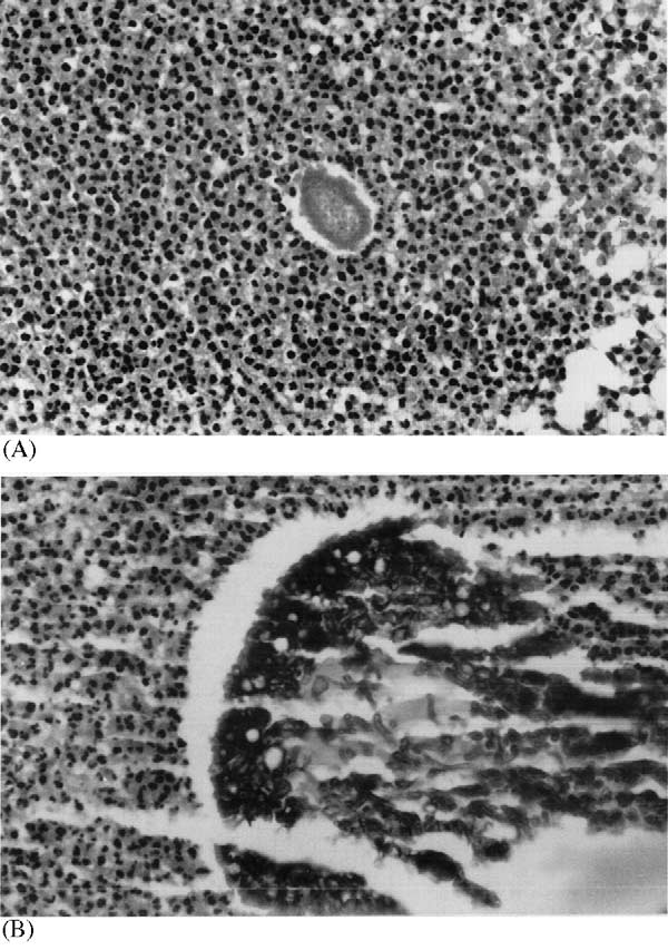
Tissue reaction pattern was similar in both groups of mycetoma. Inflammatory foci werecomposed mainly of neutrophils. The innermostneutrophils were closely attached to the surface ofthe grains. Outside this zone there was granulationtissue plasma cells and few neutrophils. Several macro-phages had large vacuolated cytoplasm. Concen-trically arranged fibrin layers, giving an onionskinappearance, surrounded capillaries and veins.
Fig. 1. (a) Actinomycetoma lesion. Neutrophils exsudate Arterioles showed hypertrophic muscular layer, around a small irregular grain bordered by short irregularspicules of eosinophilic material; (b) eumycetoma lesion. Grain thickened and edematous intima and narrowed composed of interwoven hiphae surrounded by neutrophils lumen. The dermis displayed variable degree of exsudate. Haematoxilyn and eosin ( /400 original magnifica-


C.C. Guimara˜es et al. / Acta Tropica 87 (2003) 377 /384 vesicles, sometimes embedded in a cement-like was found in both groups of mycetoma ( matrix (Five specimens exhibited dema- tiaceous hyphae.
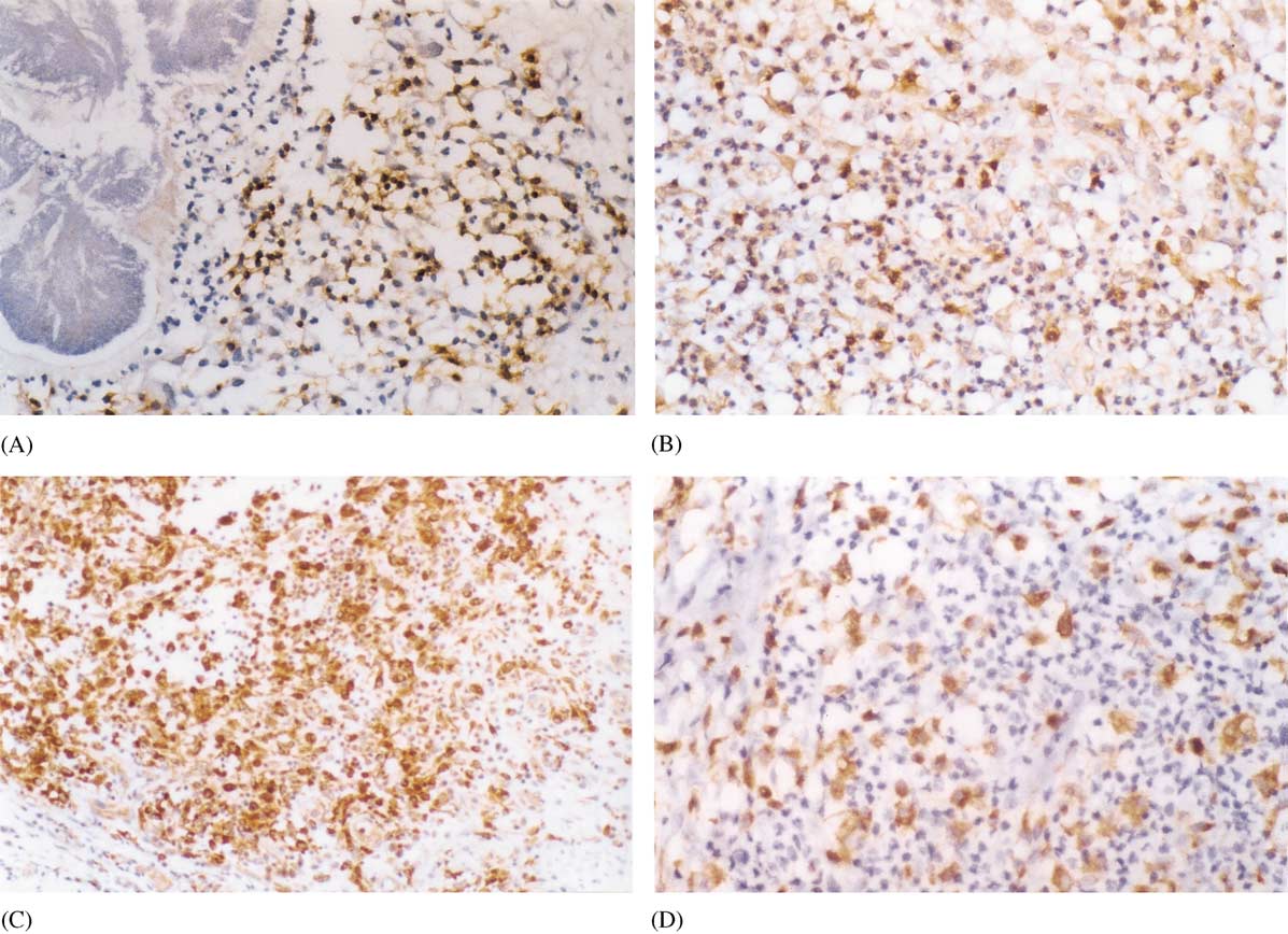
The number of CD3/ and CD4/ cells did not B lymphocytes were not seen among the inflam- differ between both groups. On the other hand, the matory cells in tissue reaction of either group. T number of CD8/ lymphocytes and macrophages lymphocytes (CD3/) were observed around the was higher in ACM lesions (P /0.02 and 0.01, neutrophils aggregates as small clusters and also respectively). shows the distribution of the interspersed among other mononuclear cells.
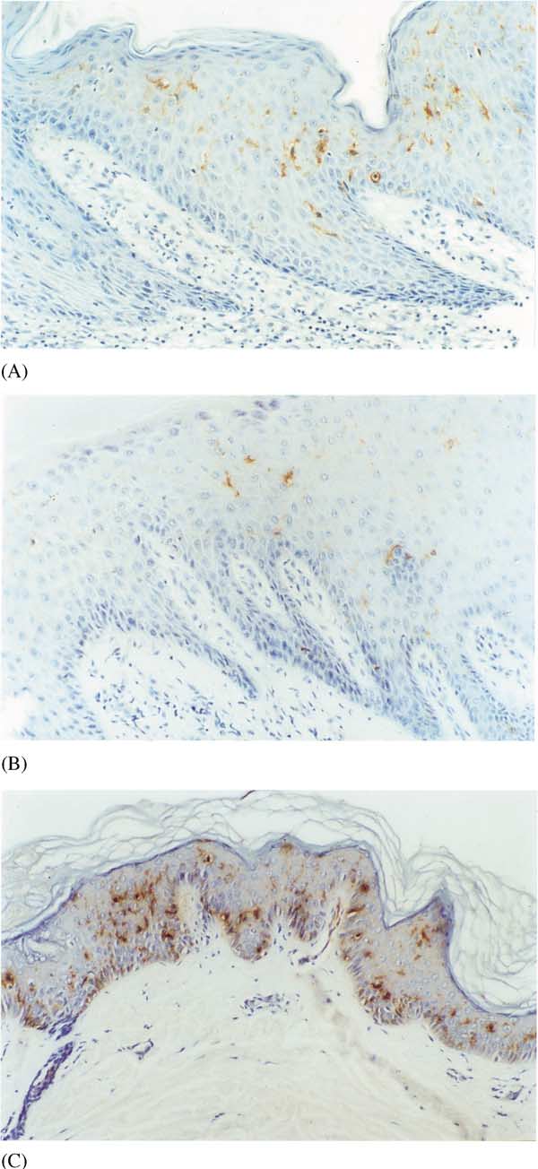
immune labeled cells/mm2 of both mycetoma CD4/ and CD8/ lymphocytes exhibited the same disposition in the inflammatory process Epidermal LC were morphologically altered, (a, b). Macrophages, with cytoplasm labeled displaying irregular and short dendritic processes.
by CD68 antibody, were observed within the When compared with the control group, both inflammatory foci. Giant multinucleated cells ACM and EUM specimens showed a statistically seen in type II reaction were strongly labeled by significant (P /0.0004) decrease in number of LC CD68 antibody. The same type of tissue reaction ). Interestingly no difference was found Fig. 2. (a) Actinomycetoma lesion. CD8/ lymphocytes around a grain ( /200 original magnification). (b) Eumycetoma lesion.
CD8/ lymphocytes intermingled with neutrophils in an eumycotic inflammatory focus ( /200 original magnification). (c)Actinomycetoma lesion. Numerous CD68/ macrophages in inflammatory focus (/200 original magnification). (d) Eumycetomalesion. CD68/ macrophages among neutrophils in the inflammatory focus ( /400 original magnification). Immunohistochemistrytechnique SABC complex.
C.C. Guimara˜es et al. / Acta Tropica 87 (2003) 377 /384 Fig. 3. Population of CD3/ lymphocytes; CD4/ and CD8/ lymphocytes and macrophages (CD68/) in the tissue reaction ofactinomycetoma (solid symbols) and eumycetoma (empty symbols). Bar-median; (a) P /0.02; (b) P /0.01 (Mann /Whitney test).
when both mycetoma groups were compared with as well as macrophages were observed around the each other (P /0.05) ( neutrophil zone and also intermingled with thesecells. ACM lesions exhibited a higher number ofCD8/ T lymphocytes and macrophages thanEUM. B lymphocytes were not seen in tissue reaction of either mycetoma group. B lymphocytesdo not participate in the so-called SALT of normal Epidemiology, clinical aspects, pathology and treatment of mycetoma have been well addressed A T lymphocyte defect has been proposed in the in medical literature ( pathogenesis of experimentally induced mycetoma Immunopathological aspects of tissue reaction ). Disseminated infection was have not been well characterized. Little is known described following Nocardia inoculation in T about the immuno-phenotype of cell elements of cell deficient mice. On the other hand, mycetoma mycetoma tissue reaction. There is only one report has been obtained by inoculation of animals addressing the phenotype of the cellular elements without lymphocyte defects in tissue reaction of ACM ( The present study demonstrates that lympho- In chromoblastomycosis, considered by cyte population in mycetoma tissue reaction is as a minimycetoma, the lesion is considered composed of both CD4 and CD8 phenotype T to be controlled by phagocytes (neutrophils and lymphocytes. Macrophages also participate in the macrophages) although this cellular defense me- inflammatory reaction. We did not observe a well chanism is inefficient in eliminating the fungi; the organized stratification of the different immuno- disease chronically evolves and is difficult to treat labeled cells in mycetoma inflammatory foci as described by . All but one T lymphocytes with a predominance of helper tissue specimen of each group exhibited type I phenotype were demonstrated in skin and lymph reaction as proposed by . T node in paracoccidioidomycosis and these cells are lymphocytes, CD4/ and CD8/ T lymphocytes, considered to be actively involved in the disease


C.C. Guimara˜es et al. / Acta Tropica 87 (2003) 377 /384 process (). T lympho-cytes with helper/inducer phenotype were also thepredominant cells observed in the site of cytoplas-mic Paracoccidioides brasiliensis antigen intrader-mal inoculation ( Actinomycotic mycetomas respond better to chemotherapy than eumycotic mycetomas (We observed that ACM lesionsexhibit a higher number of both CD8/ lympho-cytes and macrophages than EUM lesions. CD8/T lymphocytes are related to cytotoxic mechan-isms against intracellular pathogens. On the otherhand, they have been demonstrated as cytotoxic Tlymphocytes against schistosomal egg antigen andable to produce interferon gamma in a murineexperimental model (). Macro-phages secrete many cytotoxic factors whichenable them to kill parasites without ingestingthem and also to act as killer cells throughantibody dependent cell-mediated cytotoxicityThe data obtained in thepresent study might explain the better response totherapy ACM presents, i.e. a more efficaciousimmune response is present in ACM than inEUM.
The role of LC in cutaneous infectious diseases has been well studied in leishmaniasis. These cellscan be both target and nest of parasites in themurine experimental model (consideredLC as part of the protective mechanisms towardsLeishmania .
There are few papers that discuss the role of LC population in cutaneous deep mycosis. Morpho-logically altered LC, presenting short dendriticprocesses were described in paracoccidioidomyco-sis. A reduction in number of LC was also notedS100 protein positive cellswere demonstrated next to the granuloma inexperimental paracoccidioidomycosis and were Fig. 4. Epidermal LC demonstrated by CD1a antibody. (a) interpreted as participating in the process of T Actinomycetoma lesion. Note few LC with short dendrites lymphocytes activation with a possible role in the between keratinocytes in the spinous layer of epidermis ( /630 formation of granuloma ( original magnification). (b) Eumycetoma lesion. Very few LC in demonstrated an even the spinous layer of epidermis ( /630 original magnification).
(c) Control skin. Numerous LC with anastomozing dendrites distribution of S100 protein positive dendritic cells between spinous layer keratinocytes ( /400 original magnifica- in epidermis and dermis of paracoccidioidomyco- tion). Immunohistochemistry technique SABC complex.
sis skin lesions. These authors were not able todemonstrate P. brasiliensis antigen in S100/ cells C.C. Guimara˜es et al. / Acta Tropica 87 (2003) 377 /384 Fig. 5. Epidermal CD1a/ fraction of area in actinomycetoma (j), eumycetoma (') and control group (%). Bar-median; controlgroup/actinomycetoma and eumycetoma groups (P /0.004; Kruskal /Wallis test).
using the double labeling immunohistochemical Mycetoma patients are chronically exposed to technique. On the other hand, sunlight because of their job activities in rural observed the same number of CD1a areas (). Another explana- cutaneous lesions of chromomycosis and in nor- tion could be LC migration from skin to draining mal skin control group.
lymph nodes in order to promote T lymphocytes LCs play a pivotal role in the cellular immunity mechanisms. Their function includes antigen inter- We demonstrated that T lymphocytes, either nalization, processing and presentation to T lym- with CD4/ or CD8/ phenotypes, participate in phocytes. In the skin they are involved in the tissue reaction in mycetoma lesions. Although mechanisms of immune surveillance against infec- mycetoma occurs as a consequence of inoculation tious agents.
of the agent into the skin, it is not generally We have been able to demonstrate that epider- followed by systemic dissemination. T lympho- mal LC were morphologically altered and de- cytes observed in skin mycetoma foci may be creased in number in mycetoma when compared involved in the immunopathological mechanisms with normal skin, but preventing disease dissemination, although it is not observed normal number and morphology of sufficient to resolve the local infection.
epidermal LC in overlying ACM epidermis whencompared with normal skin. The cutaneous sur-face depleted of LC may allow unprocessed parasite antigens to gain access to the circulation,playing a role in the development of immune This work was supported by Conselho Nacional tolerance. The markedly reduced number of LC de Pesquisas-CNPq grant (141375/98-1). We thankCarla Pagliari and Elaine Raniero Fernandes for found in both ACM and EUM lesions may reflect immune tolerance and/or a depressed cellularimmune response secondary to the infection asproposed for paracoccidioidomycosis The degenerative aspects and LCnumerical reduction in mycetoma epidermis could Ashworth, J., Turbitt, M.L., Mackie, R., 1986. The distribution also be related to chronic exposition to ultraviolet and quantification of Langerhans cell in normal human B radiation, as described by .
epidermis. Clin. Exp. Dermatol. 11, 153 /158.
C.C. Guimara˜es et al. / Acta Tropica 87 (2003) 377 /384 Bacci, S., Romagnoli, P., Streilein, J.W., 1998. Reduction in Lacaz, C.S., 1981. Distribuic¸a˜o geogra´fica dos micetomas no number and morphologic alterations of Langerhans cells Brasil. Ann. Bras. Dermatol. 56, 167 /172.
after UVB radiation in vivo are accompanied by influx of Mahgoub, E.S., 1978. Experimental infection of athymic nude monocytoid cells into the epidermis. J. Invest. Dermatol.
New Zealand mice with mycetoma agents. Sabouraudia 16, 111, 1134 /1139.
Beaman, B.L., Scates, S.M., 1981. The role of L-forms of Mahgoub, E.S., Gumma, S.A., Hassan, A.M., 1977. Immuno- Nocardia caviae in the development of chronic mycetomas logical status of mycetoma patients. Bull. Soc. Pathol. Exo in normal and immunodeficient murine models. Infect.
70, 48 /54.
Immun. 33, 893 /907.
Marques, M., Moscardi-Bacchi, M., Marques, S., Franco, M., Bieber, T., Ruig, J., Braun-Falco, O., 1988. Comparison of 1993. Immunohistochemical characterization of mononuc- different methods for enumeration of LC in vertical lear cells in delayed hypersensivity reactions to Paracocci- cryosections of human skin. Br. J. Dermatol. 118, 385 /392.
dioides brasiliensis (paracoccidioidin test). Mycopathologia Bos, J.D., Zonneveld, I., Das, P.K., Krieg, S.R., Van Der Loos, 124, 7 /11.
C.M., Kapsenberg, M.L., 1987. The skin immune system McElroy, J.A., Prestes, C.A., Su, W.P.D., 1992. Mycetoma: (SIS): distribution and immunophenotype of lymphocyte infection with tumefaction, draining sinuses, and ‘‘grains''.
subpopulations in the normal human skin. J. Invest.
Cutis 107, 107 /110.
Dermatol. 88, 569 /573.
McGinnis, M.R., 1996. Mycetoma. Dermatol. Clin. 14, 97 / Castro, L.G.M., Belda, W., Jr, Salebian, A., Cuce´, L.C., 1993.
Mycetoma: a retrospective study of 41 cases seen in Sa˜o Moll, H., 1993. Epidermal Langerhans cells are critical for Paulo, Brazil, from 1978 to 1989. Mycoses 36, 89 /95.
immunoregulation of cutaneous leishmaniasis. Immunol.
Conde, C., Mancilla, R., Fresan, M., Ortiz-Ortiz, L., 1983.
Today 14, 383 /387.
Immunoglobulin and complement in tissues of mice infected Moscardi-Bacchi, M., Soares, A., Mendes, R., Marques, S., with Nocardia brasiliensis . Infect. Immun. 40, 1218 /1222.
Franco, M., 1989. In situ localization of T lymphocytes Dompmartin, A., Healy, A.T., Nacy, C.A., Hauser, C., subsets in human paracoccidioidomycosis. J. Med. Vet.
Meltzer, M.S., et al., 1988. Leishmania major infects and Mycol. 27 (3), 149 /158.
replicates within murine epidermal LC. J. Invest. Dermatol.
Pancre , V., Delacre, M., Herno, J., Auriault, C., 1999.
Schistosomal egg antigen-responsive CD8 T-cell population El Hassan, A.M., Fahal, A.H., Ahmed, A.O., Ismail, A., in Schistosoma mansoni infected BALB/c mice. Immunology Veress, B., 2001. The immunopathology of actinomycetoma lesions caused by Streptomyces somaliensis . Trans. R. Soc.
Roitt, I., Brostoff, J., Male, D., 1998. Immunology. In: Cell- Trop. Med. Hyg. 95, 89 /92.
Mediated Immune Reactions. Mosby International Limited, Esterre, P., Peyrol, S., Sainte-Marie, D., Pradinaud, R., London, UK, pp. 121  Grimaud, J.A., 1993. Granulomatous reaction and tissue Salinas-Carmona, M.C., 2000. Nocardia brasiliensis : from remodelling in the cutaneous lesion of chromomycosis.
microbe to human and experimental infections. Microbes Virchows Arch. A. Pathol. Anat. Histopathol. 422, 285 /291.
Infect. 2, 1373 /1381.
Fahal, A.H., Hassan, A.M., 1992. Mycetoma. Br. J. Surg. 79, Salinas-Carmona, M.C., Torres-Lopez, E., Ramos, A.I., Licon- Trillo, A., Gonzalez-Spencer, D., 1999. Immune response to Fahal, A.H., El Toum, E.A., El Hassan, A.M., Mahgoub, E.S., Nocardia brasiliensis antigens in experimental model of Gumaa, S.A., 1995. The host tissue reaction to Madurella actinomycetoma in BALB/c mice. Infect. Immun. 67, 2428 / mycetomatis : new classification. J. Med. Vet. Mycol. 33, Sandoval, M.P., Brito, T., Sotto, M.N., Santos, R.T., Franco, Gimenez, M.F., Tausk, F., Gimenez, M.M., Gigli, I., 1987.
M., 1996. Antigen distribution in mucocutaneous biopsies Langerhans' cells in paracoccidioidomycosis. Arch. Derma- of human paracoccidioidomycosis. Int. J. Surg. Pathol. 3, Holı´kova´, Z., Hercogova´, J., Plza´ki, J., Smetna, K., 2001.
Streilein, J.W., 1983. Skin-associated lymphoid tissues (SALT): Dendritic cells and their role in skin-induced immune origins and functions. J. Invest. Dermatol. 80, 12S /16S.
responses. J. Eur. Acad. Dermatol. Venereol. 15, 116  Tapia, F.J., Ca´rceres-Dittmar, G., Acun˜a, L., Mosca, W., 1989.
Hsu, S.M., Raine, L., Fanger, H., 1981. Use of avidin  Epidermal Langerhans cells in infectious diseases. Histol.
peroxidase complex (ABC) in immunoperoxidase techni- Histopathol. 4, 499 /508.
ques: a comparison between ABC and unlabeled antibody Yousif, M.A., Hay, R.J., 1987. Leucocyte chemotaxis to (PAP) procedures. J. Histochem. Cytochem. 29, 577 /580.
mycetoma agents */the effect to the antifungal drugs Key, M.E., Phillips, T., 1996. Catalysed signal amplification griseofulvin and ketoconazole. Trans. R. Soc. Trop. Med.
(CSA). An enhanced immunohistochemical staining method Hyg. 81, 319 /321.
based on biotinyl tyramide. J. NIH Res. 8, 72.
Zaias, N., 1978. Chromomycosis. J. Cutan. Pathol. 5, 155 /164.

Source: http://diagcel.com.br/pdf/Lymphocyte%20subsets,%20macrophages%20and%20Langerhans%20cells%20in%20acti.pdf

tododigital.cl

I AM A NIKON COOLPIX I AM THE NIKON COOLPIX COMPACT DIGITAL CAMERA LINEUP Viewfinder pictured on the Coolpix Capture magic moments every day 16.2 MP 7.5 cm/3-in. 18.1 MP 42x 8cm/3.2-in. 12.2 MP 5x 7.5 cm/3-in. 18.1 MP 22x 7.5 cm/3-in. DX format

Whi pharmacy manual

WHP Health Initiatives, Inc. 2275 Half Day Road, Suite 250 Bannockburn, IL 60015 WHP Health Initiatives, Inc. ("WHI") is pleased to welcome you to our network of participating pharmacies. We look forward to working with you to provide accessible, cost-effective, high quality pharmacy services for our clients and their members.