
Cialis ist bekannt für seine lange Wirkdauer von bis zu 36 Stunden. Dadurch unterscheidet es sich deutlich von Viagra. Viele Schweizer vergleichen daher Preise und schauen nach Angeboten unter dem Begriff cialis generika schweiz, da Generika erschwinglicher sind.
Home.ueb.cas.cz
The Sec6兾
8 complex in mammalian cells:
Characterization of mammalian Sec3,
subunit interactions, and expression
of subunits in polarized cells
Hugo T. Matern*, Charles Yeaman†, W. James Nelson†, and Richard H. Scheller*‡
*Genentech, Inc., Department of Richard Scheller, 1 DNA Way, South San Francisco, CA 94080-4990; and †Department of Molecular and Cellular Physiology,Stanford University Medical School, Stanford, CA 94305
This contribution is part of the special series of Inaugural Articles by members of the National Academy of Sciences elected on May 2, 2000.
Contributed by Richard H. Scheller, June 21, 2001
The yeast exocyst complex (also called Sec6兾
8 complex in higher
pattern switches from apical to isotropic the patch disperses
eukaryotes) is a multiprotein complex essential for targeting exo-
around the membrane of the bud. During cytokinesis, the
cytic vesicles to specific docking sites on the plasma membrane. It
exocyst subunits reconcentrate in a ring-like structure at the neck
is composed of eight proteins (Sec3, -5, -6, -8, -10, and -15, and
separating the mother cell and the bud. Bud tip, isotropic bud,
Exo70 and -84), with molecular weights ranging from 70 to 144
and mother–daughter neck represent sites of directed membrane
kDa. Mammalian orthologues for seven of these proteins have
growth that is coordinated with the cell cycle (1). In mammalian
been described and here we report the cloning and initial charac-
cells the sec6兾8 complex is also present on plasma membranes at
terization of the remaining subunit, Sec3. Human Sec3 (hSec3)
sites of membrane growth. In cultured hippocampal neurons, the
shares 17% sequence identity with yeast Sec3p, interacts in the
Sec6兾8 complex was shown to be present in regions of membrane
two-hybrid system with other subunits of the complex (Sec5 and
addition—i.e., at neurite outgrowth and potential active zones
Sec8), and is expressed in almost all tissues tested. In yeast, Sec3p
during synaptogenesis (9). In differentiated PC12 cells the
has been proposed to be a spatial landmark for polarized secretion
complex is found in the cell body, in the extending neurite, and
(1), and its localization depends on its interaction with Rho1p (2).
at the growth cone, whereas it shows a perinuclear localization
We demonstrate here that hSec3 lacks the potential Rho1-binding
in undifferentiated PC12 cells (10). Best characterized however
site and GFP-fusions of hSec3 are cytosolic. Green fluorescent
is the localization of the Sec6兾8 complex in Madin–Darby canine
protein (GFP)-fusions of nearly every subunit of the mammalian
kidney (MDCK) epithelial cells (8). Here the complex is rapidly
Sec6兾
8 complex were expressed in Madin–Darby canine kidney
recruited from the cytosol to cell–cell contacts on initiation of
(MDCK) cells, but they failed to assemble into a complex with
calcium-dependent cell–cell adhesion. As cell polarity develops,
endogenous proteins and localized in the cytosol. Of the subunits
the localization of the complex becomes restricted to the apical
tested, only GFP-Exo70 localized to lateral membrane sites of
junctional complex, which includes adherens junctions and tight
cell– cell contact when expressed in MDCK cells. Cells overexpress-
junctions. It has been proposed that localization of Sec6兾8
ing GFP-Exo70 fail to form a tight monolayer, suggesting the Exo70
complex to cell–cell junctions serves to direct trafficking of
targeting interaction is critical for normal development of polar-
transport vesicles containing basal-lateral proteins to the devel-
ized epithelial cells.
oping lateral membrane domain (11).
Functionally, the Sec6兾8 complex probably acts as a tethering
Vesicles mediate protein transport along the secretory path- complex at the plasma membrane. In line with the localization
way in eukaryotic cells. Transport vesicles bud from a donor
studies, it has been shown that the Sec6兾8 complex is involved
organelle and are translocated to an acceptor organelle where
in specifying docking and兾or tethering of postGolgi transport
they dock, fuse, and thereby deliver their cargo (3). Proteins that
vesicles to the plasma membrane. In yeast exocyst mutants, there
mediate different steps in vesicle trafficking are highly conserved
is an accumulation of transport vesicles in the cytoplasm, when
from yeast to man. For example, proteins that are crucial for
the cells are shifted to the restrictive temperature (12). And in
neurosecretion in mammals (nSec1, Vamp1, Vamp2, SNAP-25,
streptolysin-O permeabilized MDCK cells, antibodies to Sec8
NSF, and ␣-SNAP) are homologous to proteins required for
inhibit delivery of vesicles to the basal-lateral membrane, but not
vesicle trafficking to the yeast plasma membrane (Sec1p, Snc1p,
the apical membrane (8).
Snc2p, Sec9p, Sec18p, and Sec17p, respectively). Another group
In addition to a primary localization on the plasma membrane,
of proteins involved in this transport step in yeast includes Sec3p,
components of the exocyst complex may be present on other
Sec5p, Sec6p, Sec8p, Sec10p, Sec15p, Exo70p, and Exo84p,
membranes. Overexpressed Sec15p cofractionates in sucrose
which form a stable complex called the exocyst (4). A mamma-
gradients with Sec4p and Sncp, the rab protein, and v-SNARE
lian homolog of this protein complex (Sec6兾8 complex) has been
associated with secretory vesicles. Because Sec15p also binds to
described (5, 6), and in both yeast and mammals each subunit is
activated Sec4p, the exocyst might be an effector for this
represented once, resulting in protein complexes of 845 kDa
Rab-like GTPase that is necessary for the targeting or tethering
(yeast) and 736 kDa (rat).
of secretory vesicles to sites of secretion. Sec10p also exists in a
Accumulating evidence indicates that the Sec6兾8 complex is
free pool, as has been shown by subcellular fractionations in
required for post-Golgi vesicle trafficking (7, 8). Subcellular
yeast. Sec15p and Sec10p interact with each other in the
localization of the complex correlates with sites of polarized
two-hybrid system and
in vitro synthesized Sec15p coimmuno-
membrane growth. In yeast, Sec3p is present at plasma mem-
brane sites of active vesicle fusion, and the location of these sites
changes during the cell cycle. At the beginning of a new cell cycle,
Abbreviations: MDCK, Madin–Darby canine kidney; GFP, green fluorescent protein.
the exocyst localizes in a patch at the prebud site, and as the bud
‡To whom reprint requests should be addressed at: Genentech, Inc., 11-215, 1 DNA Way,
emerges the exocyst is localized to its tip. When the growth
South San Francisco, CA 94080-4990. E-mail:
[email protected].
9648 –9653 兩 PNAS 兩
August 14, 2001 兩 vol. 98 兩 no. 17
precipitates with epitope-tagged Sec10p (13). These findings
Sec6兾8 subunits, all possible combinations were obtained by
suggest that Sec10p and Sec15p exist in a subcomplex that might
mating Y187 (a) with AM109 (␣) and independently by cotrans-
act as a bridge between Sec4p on the vesicle and other subunits
formation of AM109 with any two plasmids. X-Gal (5-bromo-
bound to the plasma membrane.
4-chloro-3-indolyl -D-galactoside) filter assays and quantifica-
The localization of Sec3p in yeast to sites at the plasma
tion of interactions with
o-nitrophenyl--D-galactopyranoside
membrane is reported to be independent of a functional secre-
(ONPG) were performed as described (15). As negative control
tory pathway, the actin cytoskeleton, and the other exocyst
for self-activation, we used a combination of the Sec6兾8 subunits
subunits (1). This led to the model that Sec3p is a spatial
with CLONTECH vectors pGAD-T-antigen and pGBKT7-p53,
landmark for exocytosis and that it may be the component of the
while these two plasmids together served as a positive control.
complex most proximal to the target membrane. Purification of
mammalian Sec6兾8 complex hinted at the existence of a Sec3
Expression of GFP Fusion Proteins and Immunofluorescent Staining.
protein, but the corresponding gene was not previously cloned
N- and C-terminal-tagged enhanced green fluorescent fusion
(14). Coomassie blue-stained SDS兾PAGE of purified Sec6兾8
proteins were made by using pEGFP-N3 and pEGFP-C1 vectors
complex reveals eight individual bands, seven of which comprise
from CLONTECH. Specifically, we created N- and C-terminal
the components of the exocyst. But peptide sequencing of the
GFP fusion proteins of Sec3, -5, -8, and -10, and Exo70, as well
remaining protein, p106, did not easily lead to its identification
as a C-terminal Sec15-GFP fusion protein. Transfection of
in protein databases (14). Now, as whole genomes from higher
MDCK IIG epithelial cells was either performed by using
organisms are sequenced, a blast2 search with the yeast Sec3p
lipofectAMINE PLUS reagent (GIBCO兾BRL Life Technolo-
sequence lead to the identification of the
SEC3 genes from fly,
gies) or Ca2⫹-phospate. Stably transfected MDCK IIG cells
worm, and man. Here we report the cloning of the human
SEC3
expressing GFP-tagged Exo70 were selected in 500 g兾ml G418
gene, its expression pattern in different tissues, and a network of
sulfate. Stably transfected T23 MDCK cells expressing GFP-
two-hybrid interactions that link Sec3 to other subunits of the
tagged Sec3 or Sec10 under control of the tetracycline-
Sec6兾8 complex. Our localization studies employing green flu-
repressible transactivator were selected in hygromycin and main-
orescent protein (GFP) fusions of several Sec6兾8 complex
tained in DMEM containing 10% FBS and 20 ng兾ml
subunits revealed that only GFP-Exo70 becomes localized to the
doxycycline. Cells were induced to express GFP fusion proteins
plasma membrane in polarized epithelial MDCK cells, whereas
by removing doxycycline from culture medium of low-density
all other GFP-tagged subunits are cytosolic. These studies
cultures for 16–18 h before replating cells on either coverslips or
suggest that regulation of Sec6兾8 complex assembly and local-
12-mm-diameter Transwell filters (Costar). Immunofluorescent
ization at the plasma membrane depends on Exo70 targeting
staining of Sec6, E-cadherin, and ZO-1 was performed as
interactions, not Sec3 as in yeast.
described (8).
Materials and Methods
Results and Discussion
Cloning of Human SEC3. A blast2 search was done at the European
Cloning of Human SEC3. In a blast2 search at the European
Molecular Biology Laboratory (EMBL) web site, using the
Molecular Biology Laboratory (EMBL) web site, using the
entire ySec3p sequence and default parameters to identify the
entire ySec3p sequence, we identified CG3885 [DNA Data Base
human Sec3. The corresponding gene was then amplified by
in Japan (DDBJ)兾EMBL兾GenBank accession no. AAF49347],
PCR out of human cDNA (CLONTECH), using the following
F52E4.7 (accession no. T16430), and FLJ10893 (accession no.
oligonucleotide pairs and the proofreading Herculase polymer-
NP 060731兾BAA91886) as the
Drosophila,
Caenorhabditis el-
ase blend (Stratagene). HM156 (ATGACAGCAATCAAGC
egans, and human Sec3 proteins, respectively. We then cloned
the gene (AK001755) that encodes BAA91886 by PCR using
CATGTGGTTCACAGG), HM159 (GGATCAGATCTCT-
human brain cDNA as template. Stop codons in all three reading
frames upstream of the ATG start codon indicate that we indeed
CAATTAGGTTGTTCAGCTC), and HM161 (GTGGCAC-
predict the full-length protein. BAA91886 is an 894-aa protein
ACCACTGCCT GTTTCATCTGAG)兾HM165 (GGGCAAA-
with a predicted molecular weight of 101.97 kDa (GCG: mol wt).
TAAAACTGCTATATAGGTTGG). The obtained PCR prod-
The now complete calculated weight of the mammalian Sec6兾8
ucts were then cloned, sequenced, and put together by using
complex is 736 kDa and therefore roughly 110 kDa smaller then
appropriate restriction enzymes and conventional cloning.
its yeast counterpart (845 kDa). The Sec3 proteins from multi-
cellular eukaryotes share 17–22% sequence identity with the
Northern Blot Analysis. The mRNA blot of different human tissues
yeast protein. These sequence identities are low, but additional
was purchased from CLONTECH and used according to the
evidence that BAA91886 represents a bona fide ortholog of
manufacturer's instructions. The blot was probed with a 940-
ySec3p came from re-examining the peptide sequences we
nucleotide hSEC3 cDNA fragment (from bp 920 to 1860) labeled
originally reported for p106, the unidentified subunit of the
with [32P]dCTP using random primers (Megaprime RPN, Am-
Sec6兾8 complex isolated from rat brain (14). Three of these
ersham Pharmacia). To show an equal loading of RNA, the blot
peptide sequences (ELPEFNLHFF, XLQDVDLASXR, and
was stripped and tested again with human -actin cDNA as
XNRXNEPAVNVL) match (around 70% identity) within the
identified human Sec3 sequence and are preceded by lysine
residues that are recognized by trypsin to generate peptides. The
Two-Hybrid Interactions. To clone the Sec6兾8 genes into two-
deviations between the peptides and the predicted human
hybrid vectors, suitable restriction sites were created by PCR, the
sequence might be due to a combination of protein sequencing
modified regions sequenced, and the complete ORFs subse-
errors and species variations, as the peptides are derived from
quently cloned into pACTII (GAL4 activation domain vector)
rat. A sequence comparison of the yeast, fly, and human Sec3
and pGBKT7 (GAL4 DNA-binding domain vector). The two-
proteins is given in Fig. 1; the position of the three peptides is
hybrid yeast strains Y187 and AM109 were then transformed
marked by a line above the corresponding region. The newly
with these plasmids, respectively. The expression of the fusion
identified proteins are of similar length (841–894 aa) and each
protein was confirmed by Western blot analysis using antibodies
lacks ⬇480 aa found at the N terminus of the yeast protein.
directed against the HA epitope of GAL4 activation domain
Interestingly, this N-terminal domain of yeast Sec3p has recently
fusion proteins and anti c-Myc antibody to detect GAL4 DNA-
been shown to interact with Rho1p, whereas its deletion leads to
binding domain fusions. To check interactions between any two
a mislocalization of the protein (2).
Matern
et al.
PNAS 兩
August 14, 2001 兩 vol. 98 兩 no. 17 兩
9649
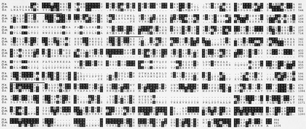
Sequence alignment of Sec3 proteins. A blast2 search with the ySec3p sequence revealed (in order): C. elegans F52E4.7, accession T16430, high score
105, e value 4.0e-08 with 19% identities; Homo sapiens BAA91886兾FLJ10893, accession NP 060731, high score 83, e value 9.0e-08 with 17% identities; andDrosophila melanogaster CG3885 protein, accession AAF49347, high score 112, e value 1.4e-06 with 22% identities. The predicted amino acid sequence of humanSec3 was compared with the respective fly and yeast homologues by using the GCG programs PILEUP and PRETTYBOX. Identical residues are in a black box with whiteletters and similar residues are shaded. Dotted regions represent gaps. Lines above the amino acid sequences indicate the peptides determined by amino-acidsequencing of the p106 subunit purified from rat brain (14). The Sec3 proteins from higher organisms lack an equivalent of the 480-aa N-terminal domain ofyeast Sec3p. Therefore, this part of ySec3p is not shown here.
Tissue Distribution of hSEC3 Transcripts. The distribution of mRNA
Molecular Interactions Between Sec6兾8 Subunits. To identify the
transcripts encoding human Sec3 was investigated by RNA
binding partners of Sec3 and those of all other subunits within
blotting (Fig. 2). The SEC3 gene is expressed as one transcript
the complex, all eight genes were subcloned into both two-hybrid
of ⬇4 kb. It is detectable in almost all tissues, but is most
vectors (pGBKT7 as bait and pACTII as prey). These constructs
abundant in brain, heart, placenta, skeletal muscle, and kidney.
were then used to test all possible pairwise interactions between
This expression pattern is similar to that of other Sec6兾8 subunits
individual subunits. An X-Gal (5-bromo-4-chloro-3-indolyl -D-
examined (6). A reblotting of the filter with a labeled probe
galactoside) filter assay that shows these interactions is given in
against -actin served as a control to show equal mRNA levels
Fig. 3A. All interactions were quantified by using a liquid
in all lines.
-galactosidase assay; but only those where the calculated in-
teraction was at least 10⫻ stronger than background (i.e., ⱖ1.0)
are given here. As shown in Fig. 3A, hSec3 interacts with Sec5
and Sec8 from rat, providing further evidence that the identified
protein is part of the Sec6兾8 complex. Other two-hybrid inter-
actions were found between Sec15 and Sec10, between Sec8 and
Sec10, between Sec5 and Sec6, and between Sec6 and Exo70. In
addition to these strong interactions, numerous weak or transient
interactions are detectable. The most plausible explanation for
these weak interactions is that the stability of the intact complex
is achieved through a series of higher order interactions not
achieved in the pairwise two-hybrid system. Intriguingly, there is
only one very strong interaction, between Sec10 and Sec15, two
proteins that in yeast have been suggested to form a subcomplex
outside the whole complex. In contrast to the other exocyst
subunits, Sec10p and Sec15p exist in a cytosolic pool, interact
with each other in the two-hybrid system, and in vitro synthesized
Sec15p coimmunoprecipitates with epitope-tagged Sec10p (13).
A schematic representation of the stronger and therefore
more reliable two-hybrid-interactions between the mammalian
Sec6兾8 subunits is given in Fig. 3B. Given the fact that both the
yeast exocyst and the mammalian Sec6兾8 complexes are com-
posed of the same number of subunits in equal stoichiometry, we
expected to find identical patterns of interactions between those
subunits. This is however only partially true. The strongest
interaction in the mammalian complex (Sec10–Sec15) was also
Multiple-tissue Northern blot analysis. Size markers are on the left in
Kb. (Upper) hSEC3, one transcript of about 4 Kb is observed in almost all
shown in yeast. The interactions between Sec6–Sec5 and Sec8–
tissues. (Lower) To show an equal loading of mRNA in all lanes, the filter was
Sec6 from rat were also seen by immunoprecipitation of in
stripped and reblotted with human -actin.
vivo-synthesized yeast exocyst proteins (13). Exo84p, however,
Matern et al.
Localization of GFP-Fusion Proteins in MDCK Cells. Because Sec3p is
proposed to serve as a spatial landmark for polarized membrane
growth in yeast (1), we were interested in determining whether
the mammalian homolog has this function as well. Therefore, we
expressed Sec3 tagged at either the N or C terminus with GFP
in MDCK cells and examined its subcellular distribution during
development of cell polarity. We reported previously that in
contact-naive MDCK cells endogenous Sec6兾8 complex is cy-
tosolic and that upon induction of E-cadherin-mediated cell–cell
adhesion the complex is recruited to lateral membrane cell–cell
contacts where it becomes assembled into a detergent-insoluble
structure (8). In both contact-naive (Fig. 4A, arrows) and
early-contacting (arrowheads) MDCK cells, GFP-tagged Sec3 is
found exclusively in the cytosol. No accumulation on plasma
membranes or other organelles was observed for Sec3, regardless
of whether the GFP tag was present at the C terminus (Fig. 4)
or the N terminus (data not shown). Sec3-GFP was cytosolic at
all time points examined over the course of 72 h following
induction of cell–cell adhesion, and was cytosolic in fully polar-
ized MDCK cells (Fig. 4B). Fractionation of MDCK cell ho-
mogenates in self-forming iodixanol density gradients confirmed
that Sec3-GFP was present almost exclusively in cytosolic frac-
tions (data not shown). Overexpression of Sec3-GFP had no
affect on recruitment of Sec6 (Fig. 4C) or the tight junction
associated protein ZO-1 (Fig. 4D) to lateral membranes. We
considered the possibility that overexpressed Sec3 fusion pro-
teins were cytosolic because limiting plasma membrane binding
sites were saturated. However, when different levels of GFP-
tagged Sec3 fusion proteins were expressed by varying the
concentration of doxycycline in the medium, Sec3 was found to
be cytosolic at even the lowest detectable expression levels.
Examination of GFP-tagged Sec3 in live MDCK cells revealed
no membrane-associated Sec3 during a 1-h imaging period (data
not shown). One plausible explanation for the failure to detect
exogenously expressed Sec3 fusion proteins on the plasma
membrane is that placement of a GFP tag at either terminus
Map of two-hybrid interactions. (A) X-Gal filter assays of all pairwise
interferes with the ability of Sec3 to associate with binding
two-hybrid interactions between individual Sec6兾8 subunits. CLONTECH vec-
partners on the membrane or within the Sec6兾8 complex. We
tors pBAD-T-antigene and pGBKT7-p53 combined with Sec6兾8 subunits served
disfavor this explanation, however, because a similarly modified
as negative control, whereas the two vectors together served as positive
Sec3p is functional and appropriately localized in yeast (1). A
control. A quantification of the two-hybrid interaction was done by a liquid
more interesting explanation is that the function of Sec3 in
o-nitrophenyl--D-galactopyranoside (ONPG) assay. The calculated units are
mammalian cells does not involve plasma membrane recruit-
given below the individual dots. Although all interactions were measured,
ment, or that its recruitment to the membrane is regulated by
only those 10⫻ higher than background (ⱖ1) are given here. (B) Schematic
mechanisms different from those that recruit other subunits of
representation of these two-hybrid interactions. Arrows indicate the direction
from bait to prey. (Known two-hybrid interactions in yeast are: Sec3p–Sec5p,
兾8 complex. One remarkable difference between Sec3
Sec5p–Sec10p, Sec10p–Sec15p, Exo84p–Sec5p, and Exo84 –Sec10p.)
proteins from different species is that an N-terminal region of
480 aa found in the yeast protein is lacking from higher
eukaryotic organisms. Recent work has shown that a region
was shown to interact with Sec5p and Sec10p in yeast (16). In the
spanning the first 320 aa of yeast Sec3p is essential for its
mammalian complex, Exo84 binds Exo70 and Sec15. Additional
interaction with Rho1p (2). When this domain is deleted, Sec3p
interactions observed between mammalian subunits that are not
no longer associates with Rho1p and is no longer restricted to
seen in yeast include Sec6 and Sec15 with Exo70, and Sec8 with
sites of polarized membrane growth. Indeed, it is then diffusely
Sec10 and vice versa. The interactions of Sec6 and Sec10 with
distributed throughout the yeast cytosol, similar to hSec3-GFP
Sec8 were confirmed recently by in vitro binding studies (10). In
expressed in MDCK cells. These results suggest that Sec3 from
Saccharomyces cerevisiae, Sec3p interacts with Sec5p and it may
higher eukaryotic cells does not recruit the other subunits to the
bind Sec6p and兾or Sec8p because, in a sec5 mutant, only Sec3p,
plasma membrane and does not function as a spatial landmark
Sec6p, and Sec8p are in the immunoisolated complex (13). In the
for secretion.
mammalian complex, Sec3 interacts with Sec5 and Sec8, which
We also examined the distribution of Sec10 tagged at its N
parallels the yeast data.
terminus with GFP (Fig. 4 E and F). In two-hybrid screens, this
Further studies are needed to better understand the organi-
subunit interacted strongly with Sec8. However, Sec10-GFP did
zation of this large complex; in particular, two-hybrid studies
not colocalize with endogenous Sec6 (Fig. 4G) or Sec8 when
need to be confirmed by other methods. As the data stand today
expressed in MDCK cells. Instead, most of the protein was
some, but not all of the interactions between components of the
cytosolic in both contact-naive (arrow) and early-contacting
two complexes are conserved between yeast and mammals. This
(arrowheads) cells (Fig. 4E). In polarized cells, a concentration
could be due to technical limitations, but may also indicate that
of Sec10-GFP was occasionally observed in a perinuclear local-
the structure (and function) of the complex in yeast and mam-
ization (Fig. 4F). Perinuclear distributions of Sec6兾8 complex
mals has likely evolved to fulfill different demands of spatial
subunits have been reported, although the precise identity of this
regulation of exocytosis in these eukaryotes.
compartment remains to be demonstrated (10, 17). As with
Matern et al.
PNAS 兩 August 14, 2001 兩 vol. 98 兩 no. 17 兩 9651
Expression of GFP-tagged Sec6兾8 complex subunits in MDCK cells. Stably transfected MDCK II cells expressing Sec3-GFP (A–D), GFP-Sec10 (E–H), or
Exo70-GFP (I–L); all GFP stainings are shown in green. Cells were fixed with 4% paraformaldehyde before extraction with 1% Triton X-100. Cells in C, G, and Jwere stained with monoclonal antibody 9H5 against endogenous Sec6, and bound antibodies were detected with Texas red donkey anti-mouse IgG. Cells in Dand H were stained with polyclonal anti-ZO-1 antibodies, which were detected with Texas red donkey anti-rabbit IgG. Cells in K and L were stained withmonoclonal antibody 3G8 against E-cadherin followed by Texas red donkey anti-mouse IgG. (Scale bars, 5 m.)
Sec3-GFP fusion proteins, overexpression of GFP-Sec10 had no
branes is in many ways different from the endogenous proteins.
affect on the recruitment and organization of endogenous Sec6
First, during early stages of cell–cell contact establishment,
(Fig. 4G) or ZO-1 (Fig. 4H) at the lateral membrane.
endogenous Sec6 is found throughout the cytosol in association
Of all of the Sec6兾8 complex subunits that we expressed as
with particulate structures and also along the entire length of
GFP fusion proteins (see Materials and Methods) in MDCK cells,
cell–cell contacts in a very fine distribution pattern (Fig. 4J). In
only Exo70-GFP was recruited to plasma membrane sites of
contrast, Exo70-GFP is not observed in the cytosol, and at
cell–cell contact (Fig. 4I). The behavior of Exo70-GFP during
cell–cell contacts it is observed in a much thicker distribution
development of cell polarity is superficially similar to what we
pattern that overlaps with that of Sec6 only in the oldest (middle)
have previously reported for endogenous Sec6 and Sec8 (8).
part of the contact (Fig. 4J, arrow). Often, very short cell–cell
However, the association of Exo70-GFP with the lateral mem-
contacts were observed that were positive for Sec6, but lacked
Matern et al.
Exo70-GFP (Fig. 4J, arrowhead). A biochemical difference was
velop a snug monolayer is further evidenced by the lower cell
also observed in the behaviors of Sec6 and Exo70-GFP. Whereas
density achieved by these cells at confluence. Although parental
extraction with Triton X-100 before fixation fails to remove Sec6
(Fig. 4L) and transfected (Fig. 4K) cells were seeded at identical
from cell–cell contact sites (8), Exo70-GFP is completely solu-
densities, the packing density of parental cells was significantly
bilized by this treatment (data not shown). Therefore, although
higher than Exo70 overexpressers. Although the ultimate height
Exo70 is recruited into developing cell–cell contact sites, this
of the cells was similar, the diameter of Exo70-GFP-transfected
subunit appears to arrive slightly later than Sec6 and Sec8, and
MDCK cells was typically 1.4⫻ that of parental cells. These
does not seem to be bound to the membrane by the same type
results show that overexpression of Exo70-GFP in MDCK cells
of interactions mediating Sec6 and Sec8 membrane association.
has dramatic effects on organization of lateral membranes and
This result suggests that individual subunits of the complex are
function of junctional complexes and demonstrate a crucial
recruited to and maintained at the membrane by different
function of Exo70 in establishing and maintaining cell–cell
mechanisms, rather then arriving there as a fully assembled
In Summary, the development of cells and tissues with spa-
In polarized MDCK cells, Exo70-GFP remains enriched along
tially organized membranes requires the polarized delivery of
lateral plasma membranes and was not observed at either apical
cargo-laden vesicles to the cell surface. A common feature of the
or basal membranes (Fig. 4K, green). However, in contrast to the
mechanism of polarized vesicle targeting in eukaryotic organ-
distributions observed for endogenous Sec6 and Sec8 (8) and
isms is the exocyst or Sec6兾8 complex, which acts as a tethering
Sec10 (18), Exo70-GFP distribution was not confined to the apex
factor at the plasma membrane for vesicles to be secreted. With
of the lateral membrane. Instead, Exo70-GFP appeared to be
this work, all eight subunits of this complex are known from yeast
to man. The function of the Sec6兾8 complex in polarized vesicle
uniformly distributed along the length of the lateral plasma
targeting is clear, yet the mechanism whereby this set of proteins
membrane in a distribution similar to that of E-cadherin (Fig. 4K,
acts is still largely unknown. Although the general mechanism of
action of the Sec6兾8 complex in yeast and mammals is likely to
Overexpression of Exo70-GFP had profound effects on the
be similar, differences revealed here may include Sec3 interac-
morphology of MDCK cells. In contrast to the crisp staining
tions with Rho1p, details of the molecular organization of the
observed for E-cadherin in parental MDCK cells (Fig. 4L),
complex, and Exo70 binding to target molecules. These and
E-cadherin staining in Exo70-GFP expressing cells was more
perhaps other mechanistic differences between yeast and mam-
diffuse, suggesting that the membranes of adjacent cells were not
malian vesicle targeting likely evolved to facilitate the more
in close proximity. Measurement of transepithelial resistances in
highly regulated development and physiology of multicellular
parental vs. Exo70-GFP-expressing clones revealed that the
organisms. Understanding the function of the exocyst or Sec6兾8
junctions between transfectants were much less tight than pa-
complex in a variety of species will therefore not only provide
rental MDCK cells. Whereas parental MDCK cells developed a
insight into the cell biology of membrane trafficking, but will also
transepithelial resistance of 252 ⍀兾cm2 24 h after plating,
lead to an appreciation of the ways in which fundamental cellular
Exo70-GFP-expressing cells had only developed a resistance of
mechanisms are modified during the evolution of complex
100 ⍀兾cm2. This failure of Exo70-GFP-expressing cells to de-
1. Finger, F. P., Hughes, T. E. & Novick, P. (1998) Cell 92, 559–571.
10. Vega, I. E. & Hsu, S. C. (2001) J. Neurosci. 21, 3839–3848.
2. Guo, W., Tamanoi, F. & Novick, P. (2001) Nat. Cell Biol. 3, 353–360.
11. Yeaman, C., Grindstaff, K. K. & Nelson, W. J. (1999) Physiol. Rev. 79,
3. Bock, J. B., Matern, H. T., Peden, A. A. & Scheller, R. H. (2001) Nature
(London) 409, 839–841.
12. Finger, F. P. & Novick, P. (1997) Mol. Biol. Cell 8, 647–662.
4. TerBush, D. R. & Novick, P. (1995) J. Cell Biol. 130, 299–312.
13. Guo, W., Roth, D., Walch-Solimena, C. & Novick, P. (1999b) EMBO J. 18,
5. Hsu, S. C., Hazuka, C. D., Roth, R., Foletti, D. L., Heuser, J. & Scheller, R. H.
(1998) Neuron 20, 1111–1122.
14. Hsu, S. C., Ting, A. E., Hazuka, C. D., Davanger, S., Kenny, J. W., Kee, Y. &
6. Kee, Y., Yoo, J. S., Hazuka, C. D., Peterson, K. E., Hsu, S. C. & Scheller, R. H.
Scheller, R. H. (1996) Neuron 17, 1209–1219.
(1997) Proc. Natl. Acad. Sci. USA 94, 14438–14443.
15. Fields, S. & Sternglanz, R. (1994) Trends Genet. 10, 286–292.
7. TerBush, D. R., Maurice, T., Roth, D. & Novick, P. (1996) EMBO J. 15, 6483–6494.
16. Guo, W., Grant, A. & Novick, P. (1999) J. Biol. Chem. 274, 23558–23564.
8. Grindstaff, K. K., Yeaman, C., Anandasabapathy, N., Hsu, S. C., Rodriguez-
17. Shin, D. M., Zhao, X. S., Zeng, W., Mozhayeva, M. & Muallem, S. (2000) J. Cell
Boulan, E., Scheller, R. H. & Nelson, W. J. (1998) Cell 93, 731–740.
Biol. 150, 1101–1112.
9. Hazuka, C. D., Foletti, D. L., Hsu, S. C., Kee, Y., Hopf, F. W. & Scheller, R. H.
18. Lipschutz, J. H., Guo, W., O'Brien, L. E., Nguyen, Y. H., Novick, P. & Mostov,
(1999) J. Neurosci. 19, 1324–1334.
K. E. (2000) Mol. Biol. Cell. 11, 4259–4275.
Matern et al.
PNAS 兩 August 14, 2001 兩 vol. 98 兩 no. 17 兩 9653
Source: http://home.ueb.cas.cz/synek/Exocyst%20-%20literatura/Matern%202001%20-%20Sec3.pdf
WARFARIN USE AMONG PATIENTS OVER THE AGE OF 65 TO TREAT ATRIAL FIBRILLATION: IS FALL RISK A FACTOR IN THE DECISION-MAKING PROCESS? A Systematic Review Presented to the faculty of the School of Nursing California State University San Marcos Submitted in partial satisfaction of the requirements for the degree of MASTER OF SCIENCE Family Nurse Practitioner
BREVE RESEÑA HISTORICA DEL DERECHO PENAL MILITAR MEXICANO Renato de J. Bermúdez Sumario: 1. Introducción al tema. 2. Siglo XIX o México Independiente: A. Etapa de 1821 a 1857.- B. La reforma.- C. Influencia doctrinal: a) Aspecto positivo.- b) Aspecto negativo.- 3. Siglo XX o México Contemporáneo: A. Primera etapa: a) Restablecimiento de la legislación


