
Cialis ist bekannt für seine lange Wirkdauer von bis zu 36 Stunden. Dadurch unterscheidet es sich deutlich von Viagra. Viele Schweizer vergleichen daher Preise und schauen nach Angeboten unter dem Begriff cialis generika schweiz, da Generika erschwinglicher sind.
Ijnmr.net2
Case Report
Chronic Osteomyelitis ina Newborn
Paediatrics Section
BaPPaditya daS, PranaB Kumar dey, SatyaBrata roy chowdhury, KalPana datta
showed features of chronic osteomyelitis. A diagnosis
A term, 14-day-old male baby was presented with high
of chronic osteomyeltis with septicaemia was made
grade fever, decreased feeding, lethargy, progressively
and treated conservatively with intravenous antibiotics
increasing swel ing and restricted movement of right
fol owed by oral. He showed good improvement. The
elbow since 7th day of life. He was born by normal vaginal
case is reported here for it's rarity as it was presented in
delivery in a hospital with uneventful antenatal, natal and
first week of life.
immediate postnatal period. X-ray of the right elbow
Keywords: Chronic osteomyelitis, Septicaemia, Neonate
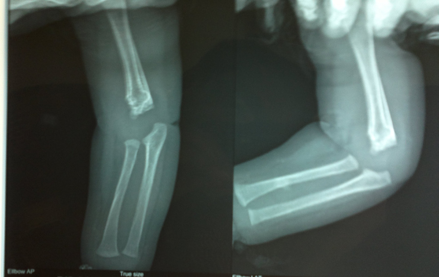
normal limit. Digital X-RAY [Table/Fig-1] of right elbow
Osteomyelitis is defined as an inflamation of the bone
(AP and Lateral view) showed metaphysical irregularity
caused by an infecting organism. It may also involve
associated with permeative destruction at the lower end
marrow, cortex, periosteum and the surrounding soft
of humeral diaphysis, continuous periosteal reaction,bony
tissues. It can be useful y sub-classified on the basis of the
fragmentation at the distomedial aspect of humeral
causative organism (pyogenic bacteria or mycobacteria),
diaphysis. All the features were suggestive of chronic
the route, duration and anatomic location of the infection
osteomyelitis. Blood culture showed no growth. Based
[1]. Osteoarticular infections, although uncommon,
on the clinical, radiological and other investigations final
represent a severe condition in neonates. To the best
diagnosis was made as chronic osteomyelitis. Baby
of our knowledge very few cases has been reported till
was managed with intravenous cefuroxime. Sling was
date. Osteoarticular, Infections in newborns are largely
maintained. After 3 weeks of intravenous therapy CRP
of an acute in nature [2]. Lower extremity joints are
was decreased, circumference came down to 12 cm
commonly affected [3]. Herein we report a rare case of
and range of movement was increased gradually. Repeat
chronic osteomyelitis of humerous since 7th day of life.
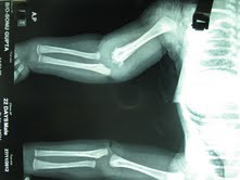
X-RAY [Table/Fig-2] showed resolution to a great extent. Baby was dischared on oral cefuroxime for three weeks
CASe RepoRT
and regular physiotherapy. After completion of antibiotic
A 14-days-old male baby was brought to us with fever,
course circumference came down to 11cm and the baby
decreased feeding, lethargy, progressively increasing
attained full range of movement of right elbow.
swelling and restricted movement of right elbow since 7th day of life. Fever was of high grade, continuous in character. Baby was born to a booked primigravida by normal vaginal delivery at term in the same institution. Antenatal and immediate post-natal periods were uneventful. Birth weight of the baby was 2.5 kg. On examination, right elbow was swollen, reddish, tender, warm to touch with circumference of 15 cm where as the left measured only 11 cm. The skin of the overlying lesion was normal.
Investigation revealed haemoglobin of 15 gm%, WBC of 34600/cmm with polymorph 75% and Platelet of
[Table/Fig-1]: X-ray of right elbow(AP & Lat view) obtained
292000/cmm. Peripheral smear showed toxic granules
on the seventh day of life at the time of admission showing
and band cell 5% of neutrophil.CRP was 9.8mg/dl and
metaphysial irregularity associated with permeative
ESR 50mm (1st hour). HIV I, II and VDRL test of the
destruction at the lower end of humeral diaphysis,continuous periosteal reaction,bony fragmentation at the
mother were negative. Coagulation profile was within
distomedial aspect of humeral diaphysis
Indian Journal of Neonatal Medicine and Research. 2013 Oct, Vol-1(2): 22-24
Bappaditya Das et al., Chronic Osteomyelitis in Newborn
cultures are positive in 30% to 50% of patients [8]. Bone aspiration under CT or ultrasound guidance may reveal an aetiologic agent when the blood cultures are negative [8]. Plain radiography is extremely helpful in diagnosis of chronic osteomyelitis which may yield areas of sequestration with dead bone lying in a pocket of cellular debris outlined by the sclerotic border, periosteal proliferative activity, modeling of the entire cortex and endosteum, and areas of bone lysis. Plain films do help in demonstrating fractures or bone malignancies, which are included in the differential diagnosis of osteomyelitis.
[9]. Technetium-labeled methylene diphosphate bone scan, CT, MRI are also useful in selective situations.
Antibiotics are recommended for at least 4–6 weeks
[Table/Fig-2]: X-ray of right elbow (AP view) showing
duration or till normalization of CRP or ESR [9]. Surgical
resolution to a great extent after 3 weeks
debridement is more critical for optimal treatment of chronic osteomyelitis [7]. Hyperbaric oxygen may be a useful adjunctive treatment measure in the management
of chronic osteomyelitis that is refractory to standard
Chronic osteomyelitis is rare in newborn. The diagnosis
approaches. So to conclude, a possible diagnosis of
is difficult and often delayed as the clinical features
osteomyelitis, though rare, should be kept in the back
differ significantly from older children, adolescent and
of mind in case of a newborn in first few weeks of life
adults [4]. In literature few cases were reported but
when there are features of inflammation and difficulty in
rarely found within first week of life. A case was reported
limb movement and a plain radiograph may be enough
where a 33 days old baby was hospitalized for pre-
to diagnose this condition in most cases.
maturity, septicemia, respiratory distress syndrome and gastrointestinal bleeding. Her chest X-ray showed
bilateral humeral osteomyelitis and bilateral glenohumeral
[1] Krogstad P. Osteomyelitis and septic arthritis. In:Feigin
RD, Cherry JD, Demmler GJ, et al, editors. Textbook of
joint arthritis [5]. Another case was reported where a five
pediatric infectious diseases. 5th edition. Philadelphia:WB
weeks old term appropriate for gestational age baby
saunders; 2004. P. 713-3
born by vacuum assisted vaginal delivery presented with
[2] Gupta V, Kumari C, Bhatia B. Chronic osteomyelitis in a
difficulty in moving his right upper limb since 3 weeks of
neonate: unusal presentation. Journal of Neonatology.
life. X ray was suggestive of osteomyelitis [3]. In a study,
2011, apr-june; 25(2):73
which included thirty-four neonates with osteomyelitis
[3] Quadir M, Ali SR, Lakhani M, Hashmi P, Amirali A. Klebsiel a
osteomyelitis of the right humerous involbing the right
showed that the hip (19 cases) was the most common
shoulder joint in an infant. J Pak Med Assoc. 2010, sept;
site involved. Swelling and pseudoparalysis were the
most significant local signs. Radiographic abnormalities,
[4] Knudsen CJM, Hoffman EF. Neonatal osteomyelitis.The
such as metaphyseal rarefaction and/or joint subluxation
Journal of bone and joint surgery.1990 sept, 72-B(5), 846-
around hip were found on the initial radiographs in
18 of the 19 cases [4]. In another study it was found
[5] Ghorashi Z, Nezami N, Hoseinpour-Feizi H, et al.
Osteomyelitis, septicaemia and meningitis caused by
that 41% cases were secondary to complication of
Klebsiella in a low birth weight newborn: a case report. J
pregnancy and 47% were secondary to complications of
Med. Case Reports 2011; 5: 241.)
deliveries. Majority of the babies had antecedent illness
[6] Weissberg Ed, Smith AL, Smith DH. Clinical features of
or were subjected to potentially infective procedures in
neonatal osteomyelitis, Pediatrics. 1974; 53: 505-10 )
perinatal period [6]. However, in our case mother had an
[7] Dubey L, Krasinski K, Hernanz-Schulman M. Osteomyelitis
secondary to trauma or infected contiguous soft tissue.
uneventful pregnancy and institutional delivery but baby
Paediatr Infect Dis J. 1988; 7:26–34.
presented with clinical features and laboratory findings
[8] Karwowska A, Davies HD, Jadavji T. Epidemiology
of of sepsis.
and outcome of osteomyelitis in the era of sequential
Chronic osteomyelitis is most commonly caused by
intravenous-oral therapy. Paediatr Infect Dis J.
S. aureus and gram-negative enterics. Polymicrobial
[9] Kaplan SL. Osteomyelitis in Children. Infect Dis Clin N Am.
aetiologies are found in a high proportion of children
19 (2005) 787–97.
with osteomyelitis secondary to trauma or contiguous spread [7]. Haematogenous spread is most common. Metaphysis of long bones, such as the femur, tibia, and humerus are usually involved [1].
The diagnosis of chronic osteomyelitis is based on clinical, laboratory and imaging studies. Standard laboratory indicators of inflammation, such as the WBC, ESR, and CRP, are all generally elevated. Blood
Indian Journal of Neonatal Medicine and Research. 2013 Oct, Vol-1(2): 22-24
Bappaditya Das et al., Chronic Osteomyelitis in Newborn
name, addreSS, e-mail id oF the
1. Dr Bappaditya Das
correSPondinG author:
2. Dr Pranab Kumar Dey
Dr Pranab Kumar Dey,
3. Dr Satyabrata Roy chowdhury
Govt. Housing Estate,Block-B, Flat-6, 82 Belgachia
4. Dr Kalpana Datta
Road,Kolkata-700037,West Bengal, India.
Phone: 918902365478
ParticularS oF contriButorS:
1. Junior Resident (MD), Department of Paediatrics,
Medical College, Kolkata, India.
Financial or other comPetinG intereStS:
2. Clinical Tutor, Department of Paediatrics, Medical
College, Kolkata, India.
3. Junior Resident (MD), Department of Paediatrics,
Medical College, Kolkata, India.
4. Professor, Department of Paediatrics, Medical
College, Kolkata, India.
Date of Publishing: oct 31, 2013
Indian Journal of Neonatal Medicine and Research. 2013 Oct, Vol-1(2): 22-24
Source: http://ijnmr.net/articles/PDF/1991/5%20-%206588_F(H)_PF1(PUH)_PFA(H)_PF1(VHP)_OLF_New.pdf
Female geneflowstratifies Hindu castesScientists have long been interested in First, there may not be enough Y-chromo- understanding how social processes some STRs to reveal a meaningful pattern, modulate evolutionary forces1. A good although a comparison of a distance matrix example of this is the intensively studied based on the Y-chromosome SNPs with the Hindu caste system, which governs the mat-
Science, Nutrition, Prévention et Santé L'artémisineUne arme contre le parasite du paludisme et les cellules cancéreuses L'artémisine est utilisée avecsuccès, seule ou associée à d'au-tres antipaludéens, pour soigner Alternatives aux statines dans le traitement le paludisme. Des rechercheseffectuées par des scientifiques


