
Cialis ist bekannt für seine lange Wirkdauer von bis zu 36 Stunden. Dadurch unterscheidet es sich deutlich von Viagra. Viele Schweizer vergleichen daher Preise und schauen nach Angeboten unter dem Begriff cialis generika schweiz, da Generika erschwinglicher sind.
Vision.seecs.edu.pk
J Med Syst (2012) 36:145–157DOI 10.1007/s10916-010-9454-7
Algorithms for the Automated Detection of DiabeticRetinopathy Using Digital Fundus Images: A Review
Oliver Faust & Rajendra Acharya U. & E. Y. K. Ng &Kwan-Hoong Ng & Jasjit S. Suri
Received: 23 January 2010 / Accepted: 28 February 2010 / Published online: 6 April 2010
# Springer Science+Business Media, LLC 2010
Abstract Diabetes is a chronic end organ disease that occurs
therefore a generalization of individual results is difficult.
when the pancreas does not secrete enough insulin or the
However, this review shows that the classification results
body is unable to process it properly. Over time, diabetes
improved has improved recently, and it is getting closer to
affects the circulatory system, including that of the retina.
the classification capabilities of human ophthalmologists.
Diabetic retinopathy is a medical condition where the retinais damaged because fluid leaks from blood vessels into the
Keywords Diabetic retinopathy . Fundus images .
retina. Ophthalmologists recognize diabetic retinopathy
Automated detection . Blood vessel area . Exudes .
based on features, such as blood vessel area, exudes,
Hemorrhages . Microaneurysms . Maculopathy
hemorrhages, microaneurysms and texture. In this paper wereview algorithms used for the extraction of these featuresfrom digital fundus images. Furthermore, we discuss
systems that use these features to classify individual fundusimages. The classifications efficiency of different DR
The fast progression of diabetes is one of the main
systems is discussed. Most of the reported systems are
challenges of current health care. The number of people
highly optimized with respect to the analyzed fundus images,
afflicted with the disease continues to grow at an alarmingrate. The World Health Organization expects the number ofpeople with diabetics to increase from 130 million to 350
O. Faust (*) R. Acharya U.
million over the next 25 years ]. The situation is made
Department of Electronics and Computer Engineering,Ngee Ann Polytechnic,
worse by the fact that only one half of the patients are
535 Clementi Road,
aware of the disease. And in the medical perspective,
Singapore, Singapore 599489
diabetes leads to severe late complications. These compli-
e-mail:
[email protected]
cations include macro and micro vascular changes which
result in heart disease, renal problems and retinopathy. For
School of Mechanical and Aerospace Engineering,
example, studies in the United States show that diabetes is
College of Engineering, Nanyang Technological University,
the fifth-deadliest disease, and still there is no cure. In the
50 Nanyang Avenue, Singapore 639798
United States, the total annual economic cost of diabetes in
2002 was estimated to be $132 billion, this translates to one
Department of Biomedical Imaging, University of Malaya,
out of every 10 health care dollars spent [
Kuala Lumpur, Malaysia
Diabetic retinopathy (DR) is a common complication of
diabetes. Indeed, it is so common that it is the leading cause
J. S. SuriBiomedical Technologies,
of blindness in the working population of western countries
The rate of diabetes is increasing, not only in developedcountries, but in underdeveloped countries as well. Unfortu-
nately, most developing countries lack basic recoding of DR
University of Idaho,Moscow, ID, USA
cases []. It is estimated that 75% of people with diabetic
J Med Syst (2012) 36:145–157
retinopathy live in developing countries []. The situation
reported that type 2 diabetes is often part of a metabolic
in developing countries is especially bad, because there is
syndrome that includes obesity, elevated blood pressure,
inadequate treatment. Regardless of the health care situation
and high levels of blood lipids ].
in their country of origin, people with diabetes are 25 timesmore likely to develop blindness when compared with
individuals who do not suffer from this disease DR isa silent disease, because it may only be recognized by the
The recent increase in diabetes can be attributed to an aging
patient when the changes in the retina have progressed to a
population and increasing prevalence of obesity as well as
level where treatment is complicated and nearly impossible.
sedentary life habits. Genetic inheritance plays a role in
The prevalence of retinopathy varies with the age of onset of
both, type 1 and type 2 diabetes. But it appears that type 1
diabetes and the duration of the disease.
diabetes is also triggered by some (mainly viral) infections.
So far, the most effective treatment for DR can be
There is also a genetic element in individual susceptibility
administered only in the first stages of the disease.
to some of these triggers which has been traced to particular
Therefore, early detection through regular screening is of
human leukocyte antigen genotypes. However, even in
paramount importance. To lower the cost of such screen-
those who have inherited the susceptibility, type 1 DM
ings, digital image capturing technology must be used,
seems to require an environmental trigger. Some evidence
because this technology enables us to employ state-of-the-
indicates that the B4 virus might be such a trigger.
art image processing techniques which automate thedetection of abnormalities in retinal images.
This paper reviews automated detection systems for DR.
This review is structured as follows: First we discuss the
Diabetes affects the kidney, eyes, nerves and heart. In the
underlying disease, i.e. diabetes, in terms of its causes and
following sections, we have discussed these affects briefly.
effects on the human body. Following the goals of thispaper, we focus on the effects of diabetes on the eye. These
Diabetic nephropathy
effects lead to features, such as blood vessel area, exudes,hemorrhages, microaneurysms and textures []. These
Diabetic nephropathy is the main cause of end-stage renal
features are used for the automatic detection of DR. In the
diseases. When the body digests protein it contaminates the
automatic detection of DR stages section we reviewed
blood with waste products. The kidneys filter out these
different automated detection systems which have been
waste products. A large number of small blood vessels
reported in scientific literature. In the discussion section we
(capillaries) are an essential component of this filter. After
discussed the advantages and disadvantages of different
20-30 years, they start to leak and useful protein is lost in
methods. The last section of this paper presents conclusions
the urine ].
and outlines further work.
It was stated that interruption of the renin–angiotensin
system slows the progression of renal diseases in patients
with type 1 diabetes, but similar data are not available forpatients with type 2 ].
Diabetes mellitus (DM) is the name of a chronic, systemic,life-threatening disease. It occurs when the pancreas does
Diabetic cardiomyopathy
not secrete enough insulin or the body is unable to processit properly. This results in an abnormal increase in the
Patients with both diabetes and ischemic heart disease seem
glucose level in the blood. Over time this high level of
to have an enhanced myocardial dysfunction leading to
glucose causes damage to blood vessels. This damage
accelerated heart failure (diabetic cardiomyopathy). Thus,
affects both eyes and nervous system, as well as heart,
patients with diabetes are prone to congestive heart failure
kidneys and other organs [].
In general there are two types of diabetes. Diabetes type
1 results from a failure of the human body to produce
Diabetic neuropathy
insulin. Type 1 diabetes is less common than type 2diabetes. People with type 1 diabetes take insulin injections.
Diabetic neuropathy results in a gradual loss of nerve
It is estimated that 90-95% of Americans, who are
function which limits the amount of sensation on the plantar
diagnosed with diabetes, have type 2 diabetes []. This
aspects of the feet [This diminished sensation disables
form of diabetes usually develops in adults age 40 and older
individuals from being able to feel the onset or occurrence
and is most common in the age group over age 55. About
of a foot injury. As a result, patients with this disease are
80% of people with type 2 diabetes are overweight. It was
more inclined to experience plantar ulceration [
J Med Syst (2012) 36:145–157
People with DM can develop nerve problems at any
following list describes three subclasses of NPDR as well
time, but the longer a person has diabetes, the greater the
risk. Acharya et al. state that abnormal plantar pressures
& Mild NPDR: at least one microaneurysm with or
play a major role in the pathologies of neuropathic ulcers in
without the presence of retinal haemorrhages, hard
the diabetic foot [].
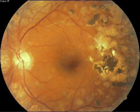
exudates, cotton wool spots or venous loops (Fig. Approximately 40% of people with diabetes have
Diabetic retinopathy
at least mild signs of diabetic retinopathy [
& Moderate NPDR: numerous microaneurysms and reti-
Diabetes mellitus often results in diabetic retinopathy which
nal haemorrhages are present. A limited amount and
is caused by pathological changes of the blood vessels
cotton wool spots of venous beading can also be seen
which nourish the retina. DR is the main cause of new cases
(Fig. 16% of the patients with moderate NPDR
of blindness among adults aged 20–74 years. During the
will develop PDR within 1 year [
first 20 years of the disease, nearly all patients with type 1
& Severe NPDR: is characterized by any one of the
diabetes and >60% of patients with type 2 diabetes have
following (4-2-1 rule) characteristics: (1) numerous
retinopathy. In the Wisconsin Epidemiologic Study of DR,
haemorrhages and microaneurysms in 4 quadrants of
3.6% of younger-onset patients (type 1 diabetes) and 1.6%
the retina (2) venous beading in 2 or more quadrants (3)
of older-onset patients (type 2 diabetes) were legally blind
Intraretinal microvascular abnormalities in at least 1
]. In the younger-onset group, 86% of blindness was
quadrant (Fig. ). Severe NPDR carries a 50%
attributable to DR. In the older-onset group, in which other
chance of progression to PDR within 1 year [
eye diseases were common, one-third of the cases of legal
& PDR: is the advanced stage; signals, sent by the retina for
blindness were due to DR. Figure shows the different
nourishment, trigger the growth of new blood vessels.
features of the typical DR image.
These blood vessels do not cause symptoms or vision loss.
DR occurs when the increased glucose level in the
But, their walls are thin and fragile, this leads to a high risk
blood damages the capillaries, which nourish the retina.
that they leak blood (Fig. ). This leaked blood
As a result of this damage, the capillaries leak blood
contaminates the vitreous gel and this causes severe
and fluid on the retina [The visual effects of this
vision loss and even blindness. About 3% of people, with
leakage are features, such as microaneurysms, hemor-
this condition, may experience severe visual loss ].
rhages, hard exudates, cotton wool spots or venous loops,of DR ].
Detection methods
Types of diabetic retinopathy DR can be broadly classi-fied as nonproliferative DR (NPDR) and proliferative
Early detection of DR is important, because treatment
DR (PDR). Depending on the presence of specific DR
methods can slow down the progression of the disease.
features, the stages can be identified , The
Most treatment methods are based on laser technology.
Fig. 1 Different features in a
J Med Syst (2012) 36:145–157
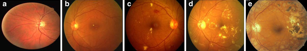
Fig. 2 Typical fundus images: (a) normal (b) mild DR (c) moderate DR (d) severe DR (e) prolific DR
Laser photocoagulation cauterizes ocular blood vessels,
provides an excellent window to the health of a patient
which effectively stops their leakage. The focal laser
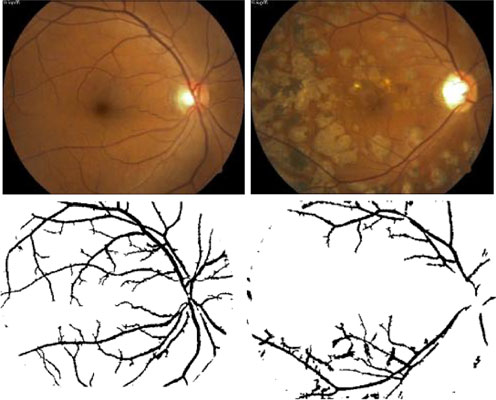
affected by DR. Figure shows an example of blood vessel
treatment method reduces retinal thickening. This may
detection from different types of DR The blood vessel
prevent worsening of retinal swelling. To be specific, this
structure was obtained by subjecting the green component
treatment reduces the risk of vision loss by 50%. For a
of the RGB fundus image to a number of image processing
small number of cases, with total vision loss, improvement
Blood vessels were detected using two-dimensional
matched filters [Gray-level profile of cross section of
blood vessel approximated by Gaussian shaped curve. Theconcept of matched filter detection of signals was used to
Medical image analysis is a research area that currently
detect piecewise linear segments of blood vessels after the
attracts lots of interest from both scientists and physicians.
The objective of this field is to develop computational tools
Vessel points in a cross section are found with a fuzzy C-
which will assist quantification and visualization of
means classifier ]. They have located and outlined blood
interesting pathology and anatomical structures. These tools
vessels in images by the use of a novel method to segment
work with digital fundus images of the eye. The procedure
blood vessels that compliments local vessel attributes with
of taking fundus images starts by dilating the pupil with
region-based attributes of the network structure.
pharmaceutical eye drops. After that the patient is asked to
Hayashi et al. have developed a computer aided diagnosis
stare at a fixation device in order to steady the eyes. While
system to assist physicians in detecting abnormalities associ-
taking the pictures, the patient will see a series of bright
ated with fundus images of the retina []. Their proposed
flashes. The entire process takes about five to ten minutes.
system can detect blood vessel intersections and it can
To ensure that DR treatment is received on time, the eye
identify abnormal widths in blood vessels.
fundus images of diabetic patients must be examined at
Computerized system for both extraction and quantita-
least once a year ].
tive description of the main vascular diagnostic signs from
Feature extraction methods and analysis
Image processing can do both reduce the workload ofscreeners and play a central role in quality assurance tasks.
Therefore, there has been an increase in the application ofdigital image processing techniques for automatic detectionof DR ]. For example, color features on Bayesianstatistical classifier were used to classify each pixel intolesion or non-lesion classes [
The following sections describe blood vessels, exudes,
hemorrhages, microaneurysms and maculopathy detectiontechniques. These detection techniques yield most of thefeatures which are used in automated DR detection systems.
Digital fundus photography from the human eye gives clearimages of the blood vessels in the retina. This method
Fig. 3 Results of blood vessel detection for normal and PDR
J Med Syst (2012) 36:145–157
fundus images in hypertensive retinopathy was presented
labeled ground truth segmentation for five images and
]. The features they have taken into account are vessel
achieved 84.37% sensitivity and 99.61% specificity.
tortuosity, generalized and focal vessel narrowing, presenceof Gunn or Salus signs.
A new system is proposed for the automatic extraction
of the vascular structure in retinal images, based on a
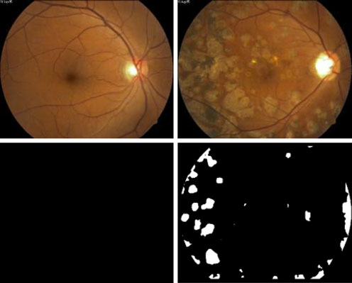
Exudates are accumulations of lipid and protein in the
sparse tracking technique was proposed . Blood vessel
retina. Typically they are bright, reflective, white or cream
points in a cross section are found by means of a fuzzy c-
colored lesions seen on the retina. They indicate increased
means classifier. After tracking the vessels, identified
vessel permeability and an associated risk of retinal edema.
segments were connected using greedy connection algo-
Although, not sight threatening in themselves, they are a
rithm. Finally bifurcations and crossings were identified
marker of fluid accumulation in the retina. However, if they
analyzing vessel end points with respect to the vessel
appear close to the macula center they are considered sight
threatening lesions. Most of the time they are seen together
Blood vessel tracker algorithm was developed to
with microaneurysms. These microaneurysms indicate
determine the retinal vascular network captured using a
themselves increased leakage, therefore the classical lesion
digital camera [The tracker algorithm detects optic
is a circular ring of exudates with several microaneurysms
disk, bright lesions such as cotton wools spots, and dark
at its center. Figure shows an example exudates detection
lesions such as haemorrhages. This algorithm identifies
from different types of DR In the result pictures, black
arteries and veins with an accuracy of 78.4% and 66.5%
indicates no exudates and white indicates the area where
exudates were detected. An important step in the extraction
Vallabha et al. have proposed a method for automated
process is removing prominent structures of the retina, such
detection and classification of vascular abnormalities in
as blood vessel tree and optic disc. After these structures
diabetic retinopathy ]. They detected vascular abnor-
have been removed, the exudates were detected using a
malities using scale and orientation selective Gabor filter
sequence of image processing algorithms
banks. The proposed method classifies retinal images as
A novel approach which combines brightness adjustment
either mild or severe cases based on the Gabor filter
procedure with statistical classification method and local-
window-based verification strategy was proposed [
The microaneurysms in retinal fluorescien angiograms
Their results indicate that they were able to achieve 100%
was identified by first locating the fovea by sub-sampling
accuracy in terms of identifying all the retinal images with
image by factor of four in each dimension ]. Subse-
exudates while maintaining a 70% accuracy in correctly
quently, the image was subjected to median filtering with a
classifying the truly normal retinal images as normal.
5 by 5 mask to reduce high-frequency components. Then
Hunter et al. have studied neural network based exudates
the image was correlated with a two-dimensional circularly
detection []. They introduced a hierarchical feature
symmetric triangular function with modelled gross shading
selection algorithm, based on sensitivity analysis to
of the macula.
Blood-vessel detection algorithm based on the regional
recursive hierarchical decomposition using quadtrees andpost-filtration of edges to extract blood vessels was studied]. This method was able to reduce false dismissals ofpredominately significant edges and faster in comparison tothe existing approach with reduced storage requirements forthe edge map.
Li et al. have used the arteriolar-to-venular diameter ratio
of retinal blood vessels as an indicator of disease relatedchanges in the retinal blood vessel tree [Theirexperimental results indicate a 97.1% success rate in theidentification of vessel starting points, and a 99.2% successrate in the tracking of retinal vessels.
A new method of texture based vessel segmentation to
overcome this problem was proposed ]. The Fuzzy C-Means (FCM) clustering algorithm was used to classify thefeature vectors into vessel or non-vessel based on thetexture properties. They compared their method with hand-
Fig. 4 Results of exudates detection for normal, PDR
J Med Syst (2012) 36:145–157
distinguish the most relevant features. The final architecture
be useful for detecting clinically important bright lesions,
achieved 91% lesion-based performance using a relatively
enhancing early diagnosis, and reducing visual loss in
small number of images.
patients with diabetes.
A new approach to automatically extract the main
A set of optimally adjusted morphological operators
features in color fundus images was proposed ]. Optic
were used for the detection of exudate in diabetic
disk was localized by the principal component analysis
retinopathy patients' non-dilated pupil and low-contrast
(PCA) and its shape was detected by a modified active
images ]. They used these operators to design an exudes
shape model (ASM). Exudates were extracted by the
detection system. This system achieved sensitivity and
combined region growing and edge detection. Their results
specificity of 80% and 99.5%, respectively.
show 99%, 94%, and 100% for disk localization, diskboundary detection, and fovea localization respectively.
Microaneurysms detection
The sensitivity and specificity for exudate detection were100% and 71%.
Microaneurysms detection is very important, because these
Osareh et al. have presented results for fundus image
structures constitute the earliest recognizable feature of DR.
based exudes classification Their method evaluated
The first reports which link these structures to DR date
different learning algorithms, such as neural network and
back to 1879 []. More recently, Jalli et al. have analyzed
support vector machine. The neural network based approach
the appearance and disappearance of microaneurysms in
performs marginally better than the support vector machine
different phases of fluorescein angiography [. In a
based approach, the latter is more flexible given boundary
similar study both formation rate and disappearance of
conditions such as control of sensitivity and specificity rates.
microaneurysms in early DR were analyzed []. The
The neural network results were: accuracy = 93.4%, sensitiv-
microaneurysms turnover were computed reliabibly from
ity = 93.0%, specificity = 94.1%.
color fundus images They used a new method called
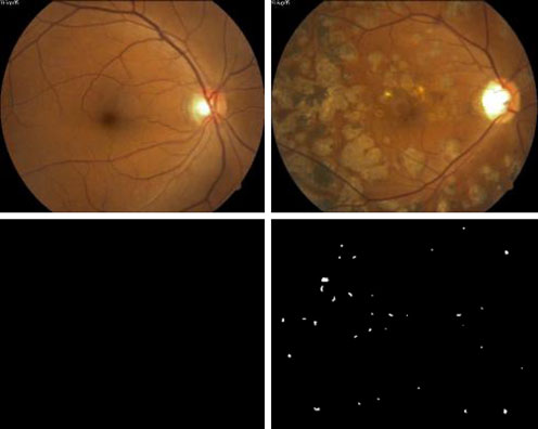
Exudates are found using their high grey level variation,
MA-tracker to count microaneurysms. They showed that
and their contours were determined by means of morpho-
the microaneurysms remain stable over time, but only 29%
logical reconstruction techniques ]. The optic disc was
remain at the same place.
detected by means of morphological filtering techniques
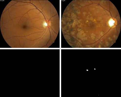
Figure shows the results of microaneurysms detection
and the watershed transformation. Their results show a
for normal and PDR []. In example the green component,
mean sensitivity of 92.8% and a mean predictive value of
of the RGB fundus image, was chosen to obtain the
microaneurysms. Similar to the exudates detection algo-
Local contrast enhancement fuzzy C-means and support
rithm, first the prominent structures within retina images,
vector machine was used to detect and classify bright
such as blood vessel tree and optic disc are to be removed.
lesions ]. Their classification results are as follows:
After that a sophisticated sequence of image processing
algorithms was used to determine the areas within the
Classification between bright lesions and bright non-
fundus images to get microaneurysms [].
lesion: sensitivity = 97%, specificity = 96%.
& Classification between exudates and cotton wool spots:
sensitivity = 88%, specificity = 84%.
Fuzzy C-means clustering and morphological reconstruc-tion was used to detect exudates detection on low-contrastimages taken from non-dilated pupils ]. The sensitivityand specificity for the exudates detection are 86% and 99%respectively.
Flemming et al. have used multi-scale morphological
algorithms to obtain what they call candidate exudates ].
The final classification was done by determining thebackground (drusen) of the candidates. Exudates weredetected with sensitivity 95.0% and specificity 84.6% in atest set of 13219 images of which 300 contained exudates.
Automated system capable of detecting exudates and
cotton-wool spots and differentiating them from drusen incolor images obtained in community based diabetic patientshas been developed ]. The machine learning can befurther improved with additional training data sets, and can
Fig. 5 Results of microaneurysms detection for normal, PDR
J Med Syst (2012) 36:145–157
The automated identification of diabetic retinopathy
exudates detection were 88.5% and 99.7%, respectively and
based on the presence of microaneurysms was studied
algorithm achieved a sensitivity of 77.5% and specificity of
]. The optometrists achieved 97 per cent sensitivity at 88
88.7% for detection of HMA.
per cent specificity and the automated retinopathy detector
Larsen et al. have used image processing for the
achieved 85 per cent sensitivity at 90 per cent specificity.
detection of both hemorrhages and microaneurysms [Their algorithm demonstrated a specificity of 71.4% and a
sensitivity of 96.7%.
The robust detection of red lesions in digital color
As the degree of DR advances retinal hemorrhages become
fundus photographs is a critical step in the development of
evident. They indicate an increased ischemia (loss of oxygen)
automated screening systems for diabetic retinopathy [
retina. As their numbers increase the retinal vessels become
Their method achieved a sensitivity of 100% at a specificity
more damaged and leaky this leads to exudation of fluid, lipid
of 87% in detecting the red lesions.
and proteins. Figure shows the result of hemorrhage
Bottom-up and top-down strategies were applied to cope
detection ]. The white patches indicates the hemorrhages
with difficulties in lesions detection, such as inhomoge-
in the image. There are two parts in haemorrhages detection:
neous illumination [After the application of appropriatestrategy, they used local contrast enhancement, fuzzy C-
i) Detection of blood vessels;
means and hierarchical support vector machine to classify
ii) Detection of blood vessels with haemorrhages.
bright non-lesion areas, exudates and cotton wool spots.
The image with blood vessel alone was subtracted fromimage with blood vessel and haemorrhages to get the image
Distance of exudates from fovea
with haemorrhages [].
Ege et al. have developed a tool which provides
In diabetic maculopathy, fluid rich in fat and cholesterol,
automatic analysis of digital fundus images ]. In their
leaks out of damaged blood vessels. If fluid and cholesterol
study, a Bayesian, a Mahalanobis, and a k nearest neighbor
accumulates near the center of the retina (the macula) it can
classifier were used on 134 retinal images. The Mahalano-
cause distortion and permanent loss of central vision. There
bis classifier showed the best results: microaneurysms,
are the two types of maculopathy eye disease:
haemorrhages, exudates, and cotton wool spots were
& Non-Clinically Significant Macular Edema (NCSME).
detected with a sensitivity of 69%, 83%, 99%, and 80%,
Figure shows a fundus image of NCSME, in this
stage the patient will not realize that he is affected,
Fully automated computer algorithms were able to detect
because there are no visible symptoms. Exudates start
hard exudates and haemorrhages and microaneurysms
to leak, and the retina becomes boggy like a sponge.
(HMA) using of a new technique, termed a 'Moat Operator',
But, the patient's vision is not seriously affected,
was proposed , The sensitivity and specificity for
because the locations of the exudates are far awayfrom the fovea.
& In the Clinically Significant Macular Edema (CSME)
stage, most of the retinal blood vessels are damaged andthe leakage area becomes bigger. The exudates leak outand this liquid concentrates very close to the fovea. Thevisibility is greatly affected, because the detected imagecannot be focused on the macula properly. Figure shows a CSME fundus image.
Fig. 6 Results of hemorrhages detection for normal, PDR []
Fig. 7 Fundus images: (a) NCSME (b) CSME
J Med Syst (2012) 36:145–157
Philips et al. have studied diabetic maculopathy and detection
aneurysms, hard exudates, and cotton wool spots, was
of exudates on fundus images [, ].
studied []. The method was able to identify the NPDR
Nayak et al. have present a computer- based system for
stage correctly with an accuracy of 81.7%.
the identification of CSME, non-CSME and normal fundus
Exudates, haemorrhages, and microaneurysms were used
eye images ]. Features are extracted from raw fundus
for screening of DR subjects [The sensitivity and
images which are then fed to an artificial neural network
specificity of their software was 74.8% and 82.7%,
classifier. They demonstrated a sensitivity of more than
respectively in differentiating DR and normal subjects
95% for these classifiers with a specificity of 100%.
Early detection of DR (presence of microaneurysms)
was proposed based on decision support system by Kahai etal. []. Bayes optimality criteria was used to detect
Texture is a measure of properties, such as smoothness,
microaneurysms. Their method was able to identify the
coarseness, and regularity of pixels, in an image ]. One
early stage of DR with a sensitivity of 100% and specificity
way to define texture is: a mutual relationship among
intensity values of neighboring pixels repeated over an area
Normal, mild, moderate, severe and prolific DR stages
larger than the size of the relationship Conventional
were automatically classified using both area and perimeter
texture recognition systems can be grouped into three
of the RGB components of the blood vessels together with
classes: structural, statistical and spectral , Textures
a feedforward neural network []. System average
can be defied using statistical approaches, this yields
classification efficiency was 84% and sensitivity, specificity
characterizations such as smooth, coarse, grainy and so
were 90% and 100% respectively. Nayak et al. have used
on. Statistical algorithms are based on the relationship
exudates and blood vessel area along with texture param-
between intensity values of pixels; measures include
eters coupled with neural network to classify fundus images
entropy, contrast, and correlation based on the gray level
into normal, NPDR and PDR [. They obtained a
co-occurrence matrix ]. Different texture parameters can
detection accuracy of 93%, sensitivity and specificity of
be used for the detection of DR stages [
90% and 100% respectively. Recently, bispectral invariantfeatures were used as features for the support vectormachine classifier to classify the fundus image in to
Automatic detection of DR stages
normal, mild, moderate, severe and prolific DR classes byAcharya et al. []. They have demonstrated an average
Over the last two decades there was a rapid development of
accuracy of 82% and sensitivity, specificity of 82% and
Computer-aided diagnosis (CAD) ]. The idea of using
88% respectively. Normal, mild, moderate, severe and
computers to help in medical image diagnosis is in more
prolific classes of DR were classified automatically based
practice. However, the quality of these CAD systems
on haemorrhages, microaneurysms, exudates and blood
increased with more accurate sensor data, more processing
vessel areas with a support vector machine classifier
power and better understanding of the underlying disease.
The system was able to identify the unknown class
Recently, Lee et al. have concluded that the performance of
accurately with an efficiency of more than 85% and a
their computer vision system in diagnosing early retinal
sensitivity of more than 82% and a specificity of 86%.
lesions is comparable with that of human experts [In
Nicolai et al. have designed an automated lesion system,
the next section we have reviewed different classification
which identified 90.1% of patients with DR and 81.3% of
patients without DR, when applied in a screening popula-tion comprising of patients with untreated DR [The
Classification methods
automated system demonstrated a sensitivity of 93.1% anda specificity of 71.6%.
Colour features were used on Bayesian statistical classifier
Usher et al. have designed a support system for DR
classify each pixel into lesion or non-lesion classes ].
screenings ]. Their system showed a maximum sensi-
They have achieved 100% accuracy in identifying all the
tivity for the detection of any retinopathy on a per patient
retinal images with exudates, and 70% accuracy in
basis of 95.1%, accompanied by a specificity of 46.3%. The
classifying normal retinal images as normal.
specificity could be increased as far as 78.9%, but this
DR and normal retina were classified automatically
increase was accompanied by a fall in sensitivity to 70.8%.
using image processing and multilayer perceptron neural
At a setting with 94.8% sensitivity and 52.8% specificity,
network ] The system yielded a sensitivity of 80.21%
no cases of sight threatening retinopathy were missed.
and a specificity of 70.66%. Automated diagnosis of
Neubauer et al. have investigated both photography and
NPDR, based on three lesions: hemorrhages and micro-
optic disc topography mode of the retinal thickness
J Med Syst (2012) 36:145–157
analyzer [The system yielded a mean 93% sensitivity
Li et al. have proposed a method for screening DR and
for PDR together with 100% specificity for DR cases.
distinguishing PDR from NPDR automatically using color
A software to grade the severity of 3 types of early
retinal images [Their method showed a sensitivity
lesions, hemorrhages and microaneurysms, hard exudates
80.5%, positive predictive value 90.8%, true positive ratio
and cotton wool spots of DR was proposed to classify
95.8%, false positive ratio 16.7% in detecting PDR and
NPDR [They were able to identify 82.6%, 82.6%, and
NPDR accurately.
88.3% using the 430 images and 85.3%, 87.5%, and 93.1%
Abràmoff et al. have evaluated the performance of a
using the 361 images, respectively, for hemorrhages and
system for automated detection of DR in digital retinal
microaneurysms, hard exudates, and cotton wool spots.
fundus images []. The system was constructed entirely
Philip et al. have assessed the efficiency of automated
from published algorithms and it was tested in a large,
"disease/no disease" grading for DR within a systematic
representative, screening population. They achieved a
screening programme [Detection of retinopathy was
sensitivity of 84% and a specificity of 64%.
achieved by automated grading with 90.5% sensitivity and
Higher order spectra features were used as input to a
67.4% specificity.
support vector machine classifier in order to classify fundus
A system, designed by Estabridis et al., has detected
images into normal, mild DR, moderate DR, severe DR and
features such as fovea, blood vessel network, optic disk,
PDR classes with an accuracy of 82%
bright and dark lesions, which are associated with DR
Vujosevic et al. have determined single lesions to grade
successfully ]. It has achieved a classification accuracy
clinical levels of DR and diabetic macular edema using
both 1 and 3 nonmydriatic digital color retinal images [
Table 1 Comparison of different classification methods
Minimum distance discriminant classifier
Sinthanayothin et
Usher et al. 2003
Singalavanija et al.
Blood vessels, exudates, haemorrhages, microaneurysms
Lee et al. 2005 []
Hemorrhages, microaneurysms, hard exudates, cotton wool spots
Neubauer et al.
Retinal thickness analyzer
Kahai et al. 2006
Decision support system (DSS)
Philip et al. 2007
Fovea, blood vessel network, optic disk, bright and dark lesions
Bright lesions, retinal vessel patterns
Abràmoff et al.
Optic disc, retinal vessels, hemorrhages, microaneurysms,
vascular, abnormalities, exudates, cotton wool spots, drusen
Area of blood vessel
Nayak et al. 2008
Blood vessels, exudates and texture
Acharya et al. 2008
Higher order spectra
Acharya et al. 2009
Blood vessel, exudates, microaneurysms, haemorrhages
Vujosevic et al.
J Med Syst (2012) 36:145–157
Sensitivity and specificity for detecting referable levels of
different texture parameters along with other DR features
DR were 82% and 92%, respectively.
can improve the classification efficiency.
Table summarizes the results of the 15 automated DR
The accurate detection of macula, optic disc, micro-
classification systems. The table entries are chronologically
aneurysm and haemorrhages is challenging. But, we feel
ordered and the percentage values for accuracy of classifi-
that, with recent advances in the medical imaging and data
cation, sensitivity and specificity are rounded to the nearest
mining techniques as well as novel algorithms for the
detection of these features, it may be possible.
Also, we feel that, the early detection of the DR (mild
DR) by detecting the microaneurysm can save the progres-
sion of the disease and hence can save the loss of visionand improve the quality of life.
The prolonged diabetes leads to the formation of micro-aneurysms and subsequently it leads to exudates as well ashaemorrhages. These are the features of DR and they may
lead to severe vision loss or even blindness. In order toavoid these complications, it is very important to detect DR
Prolonged diabetes leads to DR, where the retina is
early. This can be done by an accurate detection of
damaged due to fluid leaking from the blood vessels.
Usually, the stage of DR is judged based on blood vessels,
It is very difficult to detect the exudates clearly, because
exudes, hemorrhages, microaneurysms and texture. In this
they are tiny spots on the retina. Also, the detection of
paper, we have discussed different methods for features
haemorrhages is very challenging. The texture of haemor-
extraction and automatic DR stage detection. An ophthal-
rhages and macula is almost the same. So, we need to have
mologist uses an ophthalmoscope to visualize the blood
robust algorithms which detect these features.
vessels and his or her brain to detect the DR stages.
In the previous section we reviewed and compared 15
Recently digital imaging became available as a tool for DR
automated DR detection systems. The results were obtained
screening. It provides high quality permanent records of the
by optimizing the algorithms for a specific set of fundus
retinal appearance, which can be used for monitoring of
progression or response to treatment, and which can be
In the earlier part of the research, authors have classified
reviewed by an ophthalmologist, digital images have the
into two classes using fundus images based on two or three
potential to be processed by automatic analysis systems. A
features. Then subsequently, more features were introduced
combination of both accurate and early diagnosis as well as
to improve the classification efficiency. Also, the classifi-
correct application of treatment can prevent blindness
cation efficiency was improved further by using non-linear
caused by DR in more than 50% of all cases. Therefore,
methods like higher order spectra The algorithms
regular screenings for DR of patients with diabetes is
involving four features namely, area of blood vessel,
important. The grading of the resultant fundus images is an
exudates, haemorrhages and microaneurysms coupled with
important cost factor. Automated DR detection can reduce
support vector machine were used to classify fundus images
the grading cost and thereby make the whole screening
into five classes (normal, mild DR, moderate DR, severe
process less expensive. Some of the algorithms and systems
DR and prolific DR) with an efficiency of 86%, sensitivity
reviewed in this paper are close to achieve DR identifica-
and specificity of 82% and 86% respectively [].
tion in clinical practice.
Most of algorithms, discussed in the earlier section, have
used only a few features like blood vessels, haemorrhages,exudates and microaneurysms etc. We predict that an
algorithm involving all features namely, blood vessels,exudates, haemorrhages, microaneurysms, distance between
1. Aboderin, I., Kalache, A., Ben-Shlomo, Y., Lynch, J. W., Yajnik,
exudates and macula, and texture parameters will be more
C. S., Kuh, D., and Yach, D., Life course perspective on coronary
robust. However, for this forecast to hold it is of paramount
heart disease: key issues and implications for policy and research.
World Health Organization, Geneva, 2002.
importance that the individual parameter extraction algo-
2. Abràmoff, D. M., Niemeijer, M., Suttorp-Schulten, S. A. M.,
rithms are also as robust as possible.
Viergever, A. M., Russell, R. S., and van Ginneken, B.,
The design of good classifiers will increase the auto-
Evaluation of a system for automatic detection of diabetic
matic detection rate. Huge diverse training data will
retinopathy from color fundus photographs in a large popula-tion of patients with diabetes. Diabetes Care 31(2):193–198,
significantly improve the classification efficiency. Also,
fundus images taken under uniform good lighting con-
3. Acharya, U. R., Chua, K. C., Ng, E. Y. K., Wei, W., and Chee, C.,
ditions will improve the detection of DR. Furthermore,
Application of higher order spectra for the identification of
J Med Syst (2012) 36:145–157
diabetes retinopathy stages. J. Med. Syst., USA 32(6):431–488,
diabetic retinopathy screening. Phys. Med. Biol. 52(24):7385–
4. Acharya, U. R., Lim, C. M., Ng, E. Y. K., Chee, C., and Tamura,
22. Fong, D. S., Aiello, L., Gardner, T. W., King, G. L., Blankenship,
T., Computer based detection of diabetes retinopathy stages using
G., Cavallerano, J. D., Ferris, F. L., and Klein, R., Diabetic
digital fundus images. J. Eng. Med. 223(H5):545–553, 2009.
retinopathy. Diabetes Care 26(1):226–229, 2003.
5. Acharya, U. R., Lim, C. M., Ng, E. Y. K., Chee, C., and Tamura,
23. Forracchia, M., Grisan, M. E., and Ruggeri, A., Extraction and
T., Computer-based detection of diabetes retinopathy stages using
quantitative description of vessel features in hypertensive retinop-
digital fundus images. Proc Inst Mech Eng H. 223(5):545–553.
athy fundus images, Presented at CAFIA2001, 2001.
6. Acharya, U. R., Ng, E. Y. K., and Suri, J. S., Image modelling of
24. Frank, R. N., Diabetic retinopathy. Prog. Retin. Eye Res. 14
human eye. Artech House, MA, 2008.
7. Acharya, U. R., Tan, P. H., Subramaniam, T., Tamura, T., Chua,
25. Fujita, H., Uchiyama, Y., Nakagawa, T., Fukuoka, D., Hatanaka,
K. C., Goh, S. C., Lim, C. M., Goh, S. Y., Chung, K. R., and Law,
Y., Hara, T., Lee, G. N., Hayashi, Y., Ikedo, Y., Gao, X., and
C., Automated identification of diabetic type 2 subjects with and
Zhou, X., Computer-aided diagnosis: the emerging of three CAD
without neuropathy using wavelet transform on pedobarograph. J.
systems induced by Japanese health care needs. Comput. Methods
Med. Syst. 32(1):21–29, 2008.
Programs Biomed. 92(3):238–248, 2008.
8. Alberti, K. G., and Zimmet, P. Z., Definition, diagnosis and
26. Galloway, M. M., Texture classification using gray level run
classification of diabetes mellitus and its complications, part 1:
length. Comput. Graph. Image Process. 4:172–179, 1975.
diagnosis and classification of diabetes mellitus provisional report
27. Gonzalez, R. C., and Woods, R. E., Digital image processing, 2nd
of a WHO consultation. Diabet. Med. 15(7):539–553, 1998.
edition. Prentice Hall, New Jersey, 2001.
9. Bernardes, R., Nunes, S., Pereira, I., Torrent, T., Rosa, A., Coelho,
28. Grisan, I. E., Pesce, A., Giani, A., Foracchia, M., and Ruggeri, A.,
D., and Cunha-Vaz, J., Computer-assisted microaneurysm turn-
A new tracking system for the robust extraction of retinal vessel
over in the early stages of diabetic retinopathy. Ophthalmologica
structure, 26th Annual International Conference of the IEEE
EMBS San Francisco, USA, pp. 1620-1623, 2004.
10. Bhuiyan, A., Nath, B., Chua, J., and Kotagiri, R., Blood vessel
29. Hayashi, J., Kunieda, T., Cole, J., Soga, R., Hatanaka, Y., Lu, M.,
segmentation from color retinal images using unsupervised texture
Hara, T., and Fujita, F., A development of computer-aided
classification. IEEE Int. Conf. Image Processing, ICIP 5:521–524,
diagnosis system using fundus images, Proceeding of the 7th
International Conference on Virtual Systems and MultiMedia
11. Microaneurysms in diabetic retinopathy. Br. Med. J. 3(5774):548–
(VSMM 2001), pp. 429-438, 2001.
30. Hellstedt, T., and Immonen, I., Disappearance and formation rates
12. Brenner, M. B., Cooper, E. M., de Zeeuw, D., Keane, F. W.,
of microaneurysms in early diabetic retinopathy. Br. J. Ophthal-
Mitch, E. W., Parving, H. H., Remuzzi, G., Snapinn, M. S.,
mol. 80(2):135–139, 1996.
Zhang, Z., and Shahinfar, S., Effects of Losartan on renal and
31. Hoover, A. D., Kouzanetsova, V., and Goldbaum, M., Locating
cardiovascular outcomes in patients with type 2 diabetes and
blood vessels in retinal images by piecewise threshold probing of
nephropathy. NEJM 345(12):861–869, 2001.
a matched filter response. IEEE Trans. Med. Imag. 19(3):203–
13. Chaudhuri, S., Chatterjee, S., Katz, N., Nelson, M., and
Goldbaum, M., Detection of blood vessels in retinal images
32. Hunter, A., Lowell, J., Owens, J., and Kennedy, L, Quantification
using two-dimensional matched filters. IEEE Trans. Med. Imag.
of diabetic retinopathy using neural networks and sensitivity
analysis, In Proceedings of Artificial Neural Networks in Medicine
14. Cigna healthcare coverage position- A Report, 2007. Retrieved from:
and Biology, pp. 81-86, 2000.
33. International Council of Ophthalmology. International standards:
international clinical diabetic retinopathy disease severity scale,
Last accessed on 5th December 2007.
detailed table. Retrived from:
15. Cree, J. M., Leandro, J. J. G., Soares, J. V. B., Cesar, R. M. Jr.,
Last accessed on 17th January 2009.
Jelinek, H. F., and Cornforth, D., Comparison of various methods
34. Jalli, P. Y., Hellstedt, T. J., and Immonen, I. J., Early versus late
to delineate blood vessels in retinal images, Proceedings of the
staining of microaneurysms in fluorescein angiography. Retina 17
16th Australian Institute of Physics Congress, Canberra, 2005.
16. Diabetic Retinopathy. Retrieved from:
35. Jelinek, H. J., Cree, M. J., Worsley, D., Luckie, A., and Nixon, P.,
Last accessed on 17th January 2009.
An automated microaneurysm detector as a tool for identification
17. Early Treatment Diabetic Retinopathy Study Research Group,
of diabetic retinopathy in rural optometric practice. Clin. Exp.
Grading diabetic retinopathy from stereoscopic color fundus photo-
Optom. 89(5):299–305, 2006.
graphs: an extension of the modified Airlie House classification,
36. Kahai, P., Namuduri, K. R., and Thompson, H., A decision
ETDRS report number 10. Ophthalmology 98:786–806, 1991.
support framework for automated screening of diabetic retinopa-
18. Ege, B. M., Hejlesen, O. K., Larsen, O. V., Møller, K.,
thy. Int. J. Biomed. Imag. 2006:1–8, 2006.
Jennings, B., Kerr, D., and Cavan, D. A., Screening for
37. Kandiraju, N., Dua, S., and Thompson, H. W., Design and
diabetic retinopathy using computer based image analysis and
implementation of a unique blood vessel detection algorithm
statistical classification. Comput. Methods Programs Biomed.
towards early diagnosis of diabetic retinopathy. Proceedings of
the International Conference on Information Technology: Cod-
19. Englmeier, K. H., Schmid, K., Hildebrand, C., Bichler, S., Porta,
ing and Computing (ITCC'05) IEEE Computer Society, pp. 26-
M., Maurino, M., and Bek, T., Early detection of diabetes
retinopathy by new algorithms for automatic recognition of
38. Klein, R., Klein, B. E. K., Moss, S. E., Davis, M. D., and DeMets, D.
vascular changes. Eur. J. Med. Res. 9(10):473–488, 2004.
L., The Wisconsin Epidemiologic Study of Diabetic Retinopathy III,
20. Estabridis K, de Figueiredo RJP, Automatic detection and
prevalence and risk of diabetic retinopathy when age at diagnosis is
diagnosis of diabetic retinopathy. IEEE Int. Conf. Image Process-
30 or more years. Arch. Ophthalmol. 102(4):527–532, 1984.
ing, ICIP 2007.
39. Kulakarni, D. A., Artificial neural networks for image under-
21. Fleming, D. A., Philip, S., Goatman, A. K., Williams, J. G.,
standing. Van Nostrand Reinhold, New York, 1993. ISBN:0-442-
Olson, A. J., and Sharp, F. P., Automated detection of exudates for
J Med Syst (2012) 36:145–157
40. Kumar, A., Diabetic blindness in India: the emerging scenario.
59. Phillips, R., Spencer, T., Ross, P., Sharp, P., and Forrester, J.,
Indian J. Ophthalmol. 46(2):65–66, 1998.
Quantification of diabetic maculopathy by digital imaging of the
41. Larsen, M., Godt, J., Larsen, N., Lund-Andersen, H., Sjolie, A.
fundus. Eye 5(1):130–137, 1991.
K., Agardh, E., Kalm, H., Grunkin, M., and Owens, D. R.,
60. Ramana, K. V., and Ramamoorthy, B., Statistical methods to
Automated detection of fundus photographic red lesions in
compare the texture features of machined surfaces. Pattern
diabetic retinopathy. Invest. Ophthalmol. Vis. Sci. 44(2):761–
Recogn. 29:1447–1459, 1996.
61. Reaven, G. M., Role of insulin resistance in human disease.
42. Lee, S. C., Lee, E. T., Kingsley, R. M., Wang, Y., Russell, D.,
Diabetes 37:1595–1607, 1988.
Klein, R., and Warn, A., Comparison of diagnosis of early retinal
62. Scott, M., Grundy, C., Benjamin, I. J., Burke, G. L., Chait, A.,
lesions of diabetic retinopathy between a computer system and
Eckel, R. H., Howard, B. V., Mitch, W., Smith, S. C., and Sowers,
human experts. Arch. Ophthalmol. 119(4):509–515, 2001.
J. R., Diabetes and cardiovascular disease. A statement for
43. Lee, S. C., Lee, E. T., Wang, Y., Klein, R., Kingsley, R. M., and
Healthcare Professionals From the American Heart Association.
Warn, A., Computer classification of nonproliferative diabetic
Circulation 100:1134–1146, 1999.
retinopathy. Arch. Ophthalmol. 123(6):759–764, 2005.
63. Screening for Diabetic Retinopathy in Europe 15 years after the St.
44. Li, H., and Chutatape, O., Fundus image feature extraction.
Vincent Declaration. The Liverpool Declaration 2005. Retrieved
Proceedings 22nd Annual EMBS International Conference,
Chicago, pp. 3071-3073, 2000.
Last accessed on 20th December 2007.
45. Li, H., Hsu, W., Lee, M. L., and Wong, T. Y., Automated grading
64. Shahidi, M., Ogura, Y., Blair, N. P., and Zeimer, R., Retinal
of retinal vessel caliber. IEEE Trans. Biomed. Eng. 52:1352–1355,
thickness change after focal laser treatment of diabetic macular
oedema. Br J Ophthalmol. 78(11):827–830, 1994.
46. Li, Q., Jin, X.-M., Gao, Q., You, J., and Bhattacharya, P.,
65. Sinthanayothin, C., Boyce, J. F., Williamson, T. H., and Cook, H.
Screening diabetic retinopathy through color retinal images.
L., Automated detection of diabetic retinopathy on digital fundus
Medical Biometrics 4901:176–183, 2008.
image. Diabet. Med. 19(2):105–112, 2002.
47. Mirmehdi, M., Xian, X., and Suri, J. S., Hand book of texture
66. Sinthanayothin, C., Kongbunkiat, V., Phoojaruenchanachai, S.,
analysis. Imperial College Press, UK, 2008.
and Singalavanija, A., Automated screening system for diabetic
48. Nayak, J., Bhat, P. S., Acharya, U. R., Lim, C. M., and Kagathi,
retinopathy, 3rd international Symposium on Image and Signal
M., Automated identification of different stages of diabetic
Processing and Analysis 44(2):767-771, 2003.
retinopathy using digital fundus images. J. Med. Syst., USA, 32
67. Sopharak, A., and Uyyanonvara, B., Automatic exudates detection
from diabetic retinopathy retinal image using fuzzy C-means and
49. Nayak, J., Bhat, P. S., and Acharya, U. R., Automatic identifica-
morphological methods, Proceedings of the third IASTED
tion of diabetic maculopathy stages using fundus images. J. Med.
international conference Advances in Computer Science and
Eng. Technol. 33(2):119–129, 2009.
Technology, Thailand, pp. 359-364, 2007.
50. Neubauer, A. S., Chryssafis, C., Thiel, M., Priglinger, S., Welge-
68. Sopharak, A., Uyyanonvara, B., Barman, S., and Williamson, H.
Lussen, U., and Kampik, A., Screening for diabetic retinopathy
T., Automatic detection of diabetic retinopathy exudates from
and optic disc topography with the retinal thickness analyzer.
non-dilated retinal images using mathematical morphology meth-
Ophthalmologe 102(3):251–258, 2005.
ods. Comput. Med. Imaging Graph. 32(8):720–727, 2008.
51. Nicolai, L., Jannik, G., Michael, G., Henrik, L. A., and Michael,
69. Tan, J. H., Ng E. Y. K., and Acharya, U. R., Study of normal
L., Automated detection of diabetic retinopathy in a fundus
ocular thermogram using textural parameters. Infrared Phys.
photographic screening population. Invest. Ophthalmol. Vis. Sci.
Technol. 53(2):120–126, 2009.
70. Vallabha, D., Dorairaj, R., Namuduri, K., and Thompson, H.,
52. Niemeijer, M., van Ginneken, B., Russell, R. S., Suttorp-Schulten,
Automated detection and classification of vascular abnormalities
S. A. M., and Abramoff, D. M., Automated detection and
in diabetic retinopathy, Proceedings of 13th IEEE Signals,
differentiation of drusen, exudates, and cotton-wool spots in
Systems and Computers 2:1625-1629, 2004.
digital color fundus photographs for diabetic retinopathy diagno-
71. Vujosevic, S., Benetti, E., Massignan, F., Pilotto, E., Varano, M.,
sis. Invest. Ophthalmol. Vis. Sci. 48(5):2260–2267, 2007.
Cavarzeran, F., Avogaro, A., and Midena, E., Screening for diabetic
53. Niemeijer, M., van Ginneken, B., Staal, J., Suttorp-Schulten, M., and
retinopathy: 1 and 3 nonmydriatic 45-degree digital fundus photo-
Abramoff, M., Automatic detection of red lesions in digital color
graphs vs 7 standard early treatment diabetic retinopathy study fields.
fundus photographs. IEEE Trans. Med. Imag. 24(5):584–592, 2005.
Am. J. Ophthalmol. 148(1):111–118, 2009.
54. Ong, G. L., Ripley, L. G., Newsom, R. S., Cooper, M., and
72. Walter, T., Massin, P., Erginay, A., Ordonez, R., Jeulin, C., and
Casswell, A. G., Screening for sight-threatening diabetic retinop-
Klein, J. C., Automatic detection of microaneurysms in color
athy: comparison of fundus photography with automated color
fundus images. Med. Image Anal. 11(6):555–566, 2007.
contrast threshold test. Am. J. Ophthalmol. 137(3):445–452, 2004.
73. Wang, H., Hsu, W., Goh, K. G., and Lee, M., An effective
55. Orbis. Retrieved from: Last accessed
approach to detect lesions in colour retinal images, In Proceedings
December 2009.
of the IEEE Conference on Computer Vision and Pattern
56. Osareh, A., Mirmehdi, M., Thomas, B., and Markham, R., Compar-
Recognition, 181-187, 2000.
ative exudate classification using support vector machines and neural
74. Watkins, J. P., ABC of diabetes retinopathy. British Medical
networks, The 5th International Conf. on Medical Image Computing
Journal 326:924–926, 2003.
and Computer-Assisted Intervention, pp. 413-420, 2002.
75. Wong, L. Y., Acharya, U. R., Venkatesh, Y. V., Chee, C., Lim, C.
57. Philip, S., Fleming, A. D., Goatman, K. A., Fonseca, S.,
M., and Ng, E. Y. K., Identification of different stages of diabetic
Mcnamee, P., Scotland, G. S., Prescott, G. J., Sharp, P. F., and
retinopathy using retinal optical images. Information Sciences 178
Olson, J. A., The efficacy of automated "disease/no disease"
grading for diabetic retinopathy in a systematic screening
76. World Diabetes, A newsletter from the World Health Organiza-
programme. Br. J. Ophthalmol. 91(11):1512–1517, 2007.
tion, 4, 1998.
58. Phillips, R., Forrester, J., and Sharp, P., Automated detection and
77. Zhang, X., and Chutatape, O., Detection and classification of
quantification of retinal exudates. Graefes Arch. Clin. Exp.
bright lesions in colour fundus images. Int. Conf. on Image
Ophthalmol. 231(2):90–94, 1993.
Processing 1:139–142, 2004.
J Med Syst (2012) 36:145–157
78. Zhang, X., and Chutatape, O., Top-down and bottom-up strategies
81. Singalavanija, A., Supokavej, J., Bamroongsuk, P., Sinthanayothin,
in lesion detection of background diabetic retinopathy. IEEE
C., Phoojaruenchanachai, S., and Kongbunkiat, V., Feasibility
Computer Society Conference on Computer Vision and Pattern
study on computer-aided screening for diabetic retinopathy. Jpn. J.
Recognition 2:422–428, 2005.
Ophthalmol. 50(4):361–366, 2006.
79. Parving, H. H., Brenner, B. M., Cooper, M. E., de Zeeuw, D.,
82. Usher, D., Dumskyj, M., Himaga, M., Williamson, T. H., Nussey,
Keane, W. F., Mitch, W. E., Remuzzi, G., Snapinn, S. M., Zhang,
S., and Boyce, J., Automated detection of diabetic retinopathy in
Z., and Shahinfar, S., Effect of losartan on renal and cardiovascular
digital retinal images: a tool for diabetic retinopathy screening.
complications of patients with type 2 diabetes and nephropathy.
Diabet. Med. 21(1):84–90, 2004.
Ugeskr. Laeger 163(40):5514–5519, 2001.
83. The American Orthopaedic Foot and Ankle Society, 1999 web
80. Samuel, C. L., Elisa, T. L., Yiming, W., Ronald, K., Ronald, M.
page: (Last accessed 21.01.2010).
K., and Ann, W., Computer classification of a nonproliferative
84. Acharya, U. R., Ng, E. Y. K., and Suri, J. S., Image modeling of
diabetic retinopathy. Arch. Ophthalmol. 123:759–764, 2005.
human eye. Artech House, MA, 2008.
Source: http://vision.seecs.edu.pk/wp-content/uploads/10_springer_Algorithms-for-the-Automated-Detection-of-Diabetic-Retinopathy-Using-Digital-Fundus-Images-A-Review.pdf
La fiesta de Moros y Cristianos Puzzle de grupos 1. Formad grupos de (al menos) cinco alum- nos. Estos grupos son los grupos de base(Stammgruppen). 2. En los grupos, cada alumno/alumna escoge uno de los siguientes temas: TEMA 1: información general sobre la fiestade Moros y Cristianos en España TEMA 2: la leyenda de Alcoy
Anticoagulants in atrial fibrillation patients with chronic kidney diseaseRobert G. Hart, John W. Eikelboom, Alistair J. Ingram and Charles A. Herzog Abstract Atrial fibrillation is an important cause of preventable, disabling stroke and is particularly frequent in patients with chronic kidney disease (CKD). Stage 3 CKD is an independent risk factor for stroke in patients with atrial fibrillation. Warfarin anticoagulation is efficacious for stroke prevention in atrial fibrillation patients with stage 3 CKD, but recent observational studies have challenged its value for patients with end-stage renal disease and atrial fibrillation. Novel oral anticoagulants such as dabigatran, apixaban and rivaroxaban are at least as efficacious as warfarin with reduced risks of intracranial haemorrhage. However, all these agents undergo renal clearance to varying degrees, and hence dosing, efficacy, and safety require special consideration in patients with CKD. Overall, the novel oral anticoagulants have performed well in randomized trials of patients with stage 3 CKD, with similar efficacy and safety profiles as for patients without CKD, albeit requiring dosing modifications. The required period of discontinuation of novel oral anticoagulants before elective surgery is longer for patients with CKD owing to their reduced renal clearance. Although much remains to be learned about the optimal use of these new agents in patients with CKD, they are attractive anticoagulation options that are likely to replace warfarin in coming years.


