
Cialis ist bekannt für seine lange Wirkdauer von bis zu 36 Stunden. Dadurch unterscheidet es sich deutlich von Viagra. Viele Schweizer vergleichen daher Preise und schauen nach Angeboten unter dem Begriff cialis generika schweiz, da Generika erschwinglicher sind.
47-49.fm
pISSN 2466-1384 eISSN 2466-1392大韓獸醫學會誌 (2016) 第 56 卷 第 1 號
Korean J Vet Res(2016) 56(1) : 47 49http://dx.doi.org/10.14405/kjvr.2016.56.1.47
<Case Report>
Combination therapy of cyclosporine and prednisolone in a dog
with systemic lupus erythematosus
Yeon-Hee Kim, Min-Hee Kang, Hee-Myung Park*
Department of Veterinary Internal Medicine, College of Veterinary Medicine, Konkuk University, Seoul 05029, Korea
(Received: November 11, 2015; Revised: February 29, 2016; Accepted: March 2, 2016)
Abstract : An 11-year-old, spayed female poodle presented with fever and shifting lameness. Physical examinationrevealed hyperthermia (40.6oC), and proteinuria was detected upon urinalysis. Increased neutrophils (83%) and decreasedviscosity were revealed upon synovial fluid analysis. Serum antinuclear antibody was positive at 1 : 80. Based on thesefindings, the dog was diagnosed with systemic lupus erythematosus. Immunosuppressive therapy was initiated withprednisolone and cyclosporine, and the condition was markedly improved after the treatments. This case report describesthe clinical and laboratory findings, imaging characteristics and successful outcomes after prednisolone plus cyclosporinetherapy in a canine systemic lupus erythematosus case.
Keywords : antinuclear antibody, cyclosporine, glomerulonephritis, polyarthritis, systemic lupus erythematosus
Systemic lupus erythematosus (SLE) is a multisystemic
dal anti-inflammatory drugs. Physical examination was unre-
autoimmune disorder in which immunity is directed against
markable except for hyperthermia (40.6oC). A complete blood
various tissues or tissue components [1]. The most common
count (CBC) revealed leukocytosis (27.1 × 10³/µL; reference
clinical features include shifting lameness from polyarthritis,
interval, 5.05–16.7 × 10³/µL) with degenerative neutrophilia
ulceration of extremities caused by vasculitis, icteric and pale
(26,558/µL; reference interval, 3,000–11,000/µL). A serum
mucous membranes resulting from immune-mediated hemol-
biochemical profile revealed elevated creatine kinase (270 U/
ysis, and peripheral edema or pleural effusion due to hypoal-
L; reference interval, 100–200 U/L) and C-reactive protein
buminemia secondary to glomerulonephritis. Additionally,
(> 108 µmol/L; reference interval, < 20 µmol/L) (Table 1). A
dermatologic lesions may be present, including crusting, alope-
polymerase chain reaction (PCR) testing with a tick-borne
cia, erythema, ulceration, and hyperkeratosis [8].
disease panel including Babesia spp., Haemobartonella spp.,
Major signs of SLE are skin lesions, non-erosive polyarthri-
Anaplasma spp., Ehrlichia spp. and Borrelia burgdoferi, was
tis, hemolytic anemia, glomerulonephritis, polymyositis, leuko-
performed to rule out tick-borne diseases, and the result was
penia, and thrombocytopenia [1]. Minor signs are fever, central
negative. Radiographically, there were no evident abnormali-
nervous system symptoms, oral ulcerations, lymphadenopa-
ties on all four limbs (Fig. 1). Under general anesthesia, syn-
thy, pericarditis, and pleuritis [2]. The antinuclear antibody
ovial fluid was obtained via fine needle aspiration from
(ANA) test and lupus erythematosus (LE) cell preparations are
multiple joints (both stifles, carpi, and elbows). The fluid was
used clinically for the diagnosis of SLE, but until recently, the
transparent and had decreased viscosity. Aerobic and anaero-
ANA test is considered the most sensitive [3, 14]. Immunosup-
bic bacterial cultures of the synovial fluid were negative, and
pression is vital to treating this abnormal immune response,
the fluid cell count showed neutrophil predominance (83%).
and patients can be treated with high dose of prednisolone (1–
The results characterized an inflammatory arthropathy (Fig.
2 mg/kg, per oral [PO], q12h) and cytotoxic drugs, such as
2), and the ANA test was positive (1/80). Moderate pro-
azathioprine, cyclosporine, and cyclophosphamide [4, 5]. This
teinuria (100 mg/dL) without urinary tract infection was
case report describes successful treatment using prednisolone
detected on urine dipstick and sediment examination. Uri-
and cyclosporine in a dog with SLE.
nary specific gravity was 1.025 (Table 2). Both aerobic and
An 11-year-old, 6.1 kg, spayed female poodle dog pre-
anaerobic bacterial cultures from the urine sample were per-
sented with fever, lethargy, anorexia, and shifting lameness.
formed, and the results were negative. Therefore, the pro-
The dog had a history of reluctance to stand up and walk,
teinuria was caused by glomerular damage, not lower urinary
which was intermittent and partially responsive to non-steroi-
*Corresponding authorTel: +82-2-450-4140,
Fax: +82-2-444-4396
Yeon-Hee Kim, Min-Hee Kang, Hee-Myung Park
Table 1. A complete blood count and serum biochemical results in a dog with systemic lupus erythematosus
Reference interval
Creatinine (mg/dL)
Total protein (g/dL)
Creatine kinase (U/L)
C-reactive protein (µmol/L)
D, days after first examination; WBC, white blood cells; RBC, red blood cells; PCV, packed cell volume; Hb, hemoglobin; PLT, platelets;ALT, alanine aminotransferase; AST, aspartate aminotransferase; ALP, alkaline phosphatase; BUN, blood urea nitrogen; ND, not determined.
Table 2. Urinalysis in a dog with systemic lupus erythematosus
Reference interval
USG, urine specific gravity; UPCR, urine protein creatinine ratio. *5–10 cells/µL. †100 mg/dL. ‡30 mg/dL.
Fig. 1. Radiographs of four limbs in a dog with systemic lupus
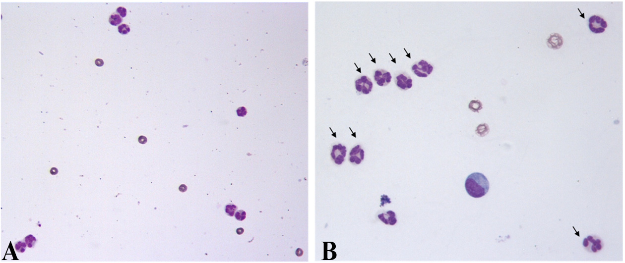
Fig. 2. Synovial fluid from the left stifle of a dog with systemic
erythematosus. No evidence of bone density loss was found in the
lupus erythematosus. Nucleated cell counts were increased (A),
limbs. (A) Left stifle joint. (B) Right stifle joint. (C) Both carpal
and non-degenerative neutrophils (B, arrows) showed predom-
inance (83%). Diff-Quik stain. 400× (A) and 630× (B).
This dog satisfied the criteria for a definite SLE condition,
with two major signs (polyarthritis and glomerulonephritis)
detected on the 15th day after treatment began. These
and a positive ANA assay; therefore, a diagnosis of definite
changes suggested secondary hepatocellular damage due to
SLE was made. Prednisolone (1 mg/kg, PO, q12h; Yuhan,
prednisolone administration. To prevent further hepatocellu-
Korea) and cyclosporine (8 mg/kg, PO, q24h; Novatis, Swit-
lar damage, a liver protectant (Zentonil 0.1 T/kg divided;
zerland) were initiated, and the dog's clinical signs including
Vétoquinol, France) was additionally prescribed. As the dos-
lameness, fever, and anorexia were improved over the next
age of prednisolone administration was tapered every two
two days. However, markedly increased liver enzymes were
weeks, the liver enzymes also decreased.
Combination therapy of systemic lupus erythematosus
The patient was monitored every 15 days, including physi-
kg, PO, q12h), but proteinuria was not controlled [9].
cal examination, CBC, serum biochemical profile, urinaly-
The patient in the current case received treatment with
sis, and urine protein creatinine ratio (UPCR). The clinical
cyclosporine and prednisolone for definite SLE. Cyclospo-
signs improved rapidly with this treatment, whereas the C-
rine was considered first in this case since the main side
reactive protein levels and UPCR gradually improved over a
effect of azathioprine is myelosuppression. Because combi-
period of months (Tables 1 and 2). Plasma cyclosporine con-
nation therapy of cyclosporine and prednisolone was applied
centration reached the therapeutic level (310 ng/mL; thera-
in the case, the dose of prednisolone used was much lower
peutic range, 100–500 ng/mL) two weeks after treatment. The
than was reported in previous case studies. The clinical signs
initial dosage of prednisolone was 1 mg/kg, PO, q12h for 15
improved, and recurrence of the condition was not observed.
days, which was then reduced by half every 15 days. Finally,
This case showed a diagnosis of definite SLE in a dog that
the prednisolone medication was discontinued 60 days after
satisfied the criteria of two major signs with a positive ANA
the first treatment. Plasma cyclosporine concentration was
test result. Unlike in previous treatment results, improve-
monitored on days 30 and 60, and reached therapeutic levels
ment of proteinuria was marked with the administration of
each time. The dosage of cyclosporine (8 mg/kg, PO, q24h)
prednisolone with cyclosporine in this dog. Thus, the use of
was maintained depending on the dog's response and her
prednisolone and cyclosporine is worthwhile to try in SLE
plasma cyclosporine concentration. No recurrence of the con-
patients with proteinuria.
dition was observed during three months of follow-up.
In conclusion, definite SLE with proteinuria was well-con-
SLE is characterized by a broad spectrum of clinical symp-
trolled by using prednisolone and cyclosporine in this dog.
toms and a multitude of laboratory abnormalities [3, 5]. Thediagnosis of SLE can be described as ‘definite SLE' or
‘probable SLE', depending on specific criteria [2]. A diagno-sis of definite SLE can be made either if a positive ANA titer
1. Bennett D. Immune-based non-erosive inflammatory joint
or LE cell preparation is identified in conjunction with two
disease of the dog. 1. Canine systemic lupus erythematosus.
major signs, or if two minor signs and one major sign are
J Small Anim Pract 1987, 28, 871-889.
identified along with a positive ANA assay or LE cell prepa-
2. Berent A, Cerundolo R. Systemic lupus erythematosus.
Compend Contin Educ Prac Vet 2005,
ration [1, 8, 14]. A probable SLE diagnosis can be made
3. Bonagura JD, Twedt DC. Kirk's Current Veterinary Therapy
either if a positive ANA titer or LE test is identified in con-
XV. pp. 268-274, Elsevier Saunders, St. Louis, 2013.
junction with one major sign, or if two major signs and a
4. Clements DN, Gear RN, Tattersall J, Carmichael S, Bennett
negative ANA titer or LE test are identified [5]. In our coun-
D. Type I immune-mediated polyarthritis in dogs: 39 cases
try, several patients were diagnosed with SLE. There were
(1997-2002). J Am Vet Med Assoc 2004, 224, 1323-1327.
two cases of definite SLE [9, 11] and one case of probable
5. Ettinger SJ, Feldman EC. Textbook of Veterinary Internal
SLE [11]. One patient with definite SLE presented with
Medicine. 7th ed. pp. 783-788, Elsevier Saunders, St. Louis,
thrombocytopenia, polyarthritis, and a positive LE cell prep-
aration [11]. The other definite SLE patient showed skin
6. Feutren G. Cyclosporin A: recent developments in the
lesions, polyarthritis, proteinuria, a positive ANA test, and a
mechanism of action and clinical application. Curr Opin
positive LE cell preparation [9].
Immunol 1989, 2, 239-245.
For the treatment of SLE, prednisolone is preferred for
Harley L, Langston C. Proteinuria in dogs and cats. Can
Vet J 2012, 53, 631-638.
immune suppression, while cyclophosphamide, cyclosporine,
8. Jones DR. Canine systemic lupus erythematosus: new insights
and levamisole are considered as alternatives [5, 10]. Cyclos-
and their implications. J Comp Pathol 1993, 108, 215-228.
porine is primarily an immunosuppressive drug that selec-
9. Kim J, Kim K, Ko I, Lee K, Na K, Yang M. A case of
tively and reversibly inhibits only the T cell-mediated response
systemic lupus erythematosus in a dog. Korean J Vet Clin
[6]. In the human literature, the effects and safety of cyclos-
Med 2000, 17, 443-449.
porine in SLE have been reported [13]. In the veterinary lit-
10. Krüger RM, França RT, Amaral AS, Schossler JEW.
erature, cyclosporine has been shown to effectively control
Polyarthritis due to systemic lupus erythematosus in a dog.
proteinuria that is refractory to steroids [3, 12]. Cyclosporine
Arq Bras Med Vet Zootec 2013, 65, 393-396.
can also help in treating proteinuria in nephropathy [7].
11. Lee CW, Na KJ, Lim JS, Seo JW. Systemic lupus
The two above-mentioned definite SLE patients were
erythematosus in a dog, suspected systemic lupus erythematosusin a dog, and autoimmune thrombocytopenic purpura hemorrha-
treated with prednisolone and azathioprine [11] or predniso-
gica in a dog. Korean J Vet Clin Med 1996, 13, 81-86.
lone monotherapy [9]. In the first patient, the clinical signs
12. Littman MP. Protein-losing nephropathy in small animals.
recurred with prednisolone (5 mg/dog, PO, q12h) alone and
Vet Clin North Am Small Anim Pract 2011, 41, 31-62.
appeared iatrogenic hyperadrenocorticism due to administra-
13. Manger K, Kalden J, Manger B. Cyclosporin A in the
tion of prednisolone. Azathioprine (11.4 mg/dog, PO, q12h)
treatment of systemic lupus erythematosus: results of an open
was added, but the clinical signs were not completely con-
clinical study. Br J Rheumatol 1996, 35, 669-675.
trolled [11]. The skin lesions and clinical symptoms of arthri-
14. Tizard IR. Veterinary Immunology. 9th ed. pp. 423-428,
tis in the second patient recovered with prednisolone (2 mg/
Elsevier, St. Louis, 2012.
Source: http://kjvr.org/upload/2016/03/30/20160330162304400140.pdf
Plasma Chem Plasma Process (2012) 32:165–176DOI 10.1007/s11090-011-9336-x Effect of Dielectric Barrier Discharge Treatmentof Blood Plasma to Improve Rheological Propertiesof Blood Jin M. Jung • Yong Yang • Dong H. Lee • Greg Fridman •Alexander Fridman • Young I. Cho Received: 19 August 2011 / Accepted: 28 November 2011 / Published online: 4 December 2011Ó Springer Science+Business Media, LLC 2011
CENTER OF SPECIAL TELECOMMUNICATIONS CYBER SECURITY CENTER CERT-GOV-MD BE WARNED, STAY PROTECTED. November 2014 Newsletter Dear Colleagues, Cyber Security Center CERT-GOV-MD is glad to announce its newsletter, as part of its proactive services. This newsletter compiles events of IT security for


