Untitled

European Journal of Echocardiography (2009) 10, 194–212doi:10.1093/ejechocard/jep005 EAE RECOMMENDATIONS Contrast echocardiography: evidence-basedrecommendations by European Associationof Echocardiography Roxy Senior1*, Harald Becher2, Mark Monaghan3, Luciano Agati4, Jose Zamorano5,Jean Louis Vanoverschelde6, and Petros Nihoyannopoulos7 1Department of Cardiology, Northwick Park Hospital, Imperial College, London, Harrow HA1 3UJ, UK; 2John RadcliffeHospital, Oxford, UK; 3King's College Hospital, London, UK; 4La Sapienza University, Rome, Italy; 5Hospital Clı´nico San Carlos,Madrid, Spain; 6Cliniques Universitaires St-Luc, Universite ´ Catholique de Louvain, Brussels, Belgium; and 7Hammersmith Hospital, Imperial College, London, UK Received 30 December 2008; accepted 11 January 2009 This paper examines the evidence for contrast echocardiography, both for improving assessment of left ventricular structure and function compared with unenhanced echocardiography and for the identifi- cation of myocardial perfusion. Based on the evidence, recommendations are proposed for the clinicaluse of contrast echocardiography.
(which has relatively lower myocardial blood volume).
With the advancement in ultrasound techniques and In a significant proportion of patients, echocardiography improved microbubble technology, it is now possible also fails to produce diagnostically useful images despite tissue to assess myocardial microcirculation and hence perfusion.
harmonic imaging.1 The main impediments appear to be This paper examines the clinical efficacy and safety of obesity and lung disease.2 The problem is even greater in ultrasound contrast agents and proposes evidence-based patients referred for stress echocardiography as images recommendations and protocols for the use of contrast are suboptimal in as many as 33% of the patients.3 This echocardiography in various clinical scenarios.
results in inaccurate assessment of left ventricular (LV) func-tion, and suboptimal reproducibility especially in the sub-group subsequent referrals for other tests because of uninterpre- Echogenicity and ultrasound properties are determined by table images.
the size, shell, and encapsulated gas characterizing the These concerns prompted the facilitation of contrast microbubbles within the various contrast agents. Micro- ultrasound imaging that utilizes contrast agents. The bubble ultrasound scatter is proportional to the sixth present generation of ultrasound contrast agents consist of power of the radius, so the largest bubble capable of microbubbles of encapsulated high-molecular-weight gas.
passing through the pulmonary microcirculation will have Since the ultrasound characteristics of microbubbles are dis- the best acoustic profile.4 The harmonic properties of micro- tinctly different from those of the surrounding blood cells bubbles are a function of their non-linear oscillation, which and cardiac tissue, the backscatter that they produce means that they reflect sound not only at the fundamental result in intense echocardiographic signals, which are pro- frequency of the ultrasound source but also at higher harmo- portional to the blood volume. Thus, the LV cavity is nics.5 A microbubble's ultrasonic characteristics also depend enhanced compared with the surrounding heart muscle on its size, the composition of the outer shell of the bubble,and the encapsulated gas.5 In general, the more elastic theshell, the more easily it will be compressed in an ultrasonic * Corresponding author. Tel: þ44 20 8869 2547/2548; fax: þ44 20 8864 field and the better it will resonate. Conversely, stiffer E-mail address: [email protected] Published on behalf of the European Society of Cardiology. All rights reserved. & The Author 2009.
For permissions please email: [email protected].
Evidence-based recommendations by EAE on contrast echocardiography Characteristic of currently available contrast agents in UK and Europe Sulphur hexafluoride Naturally occurring lipids Contraindications and precautionsPatients experiencing side effects 11 in clinical trials (%) Most frequent side effects in Headache (2.1%), nausea (1.3%), Headache (5.4%), nausea and/ Headache (2.0%), flushing (1.0%), chest pain (1.3%), taste or vomiting (4.3%), warm back pain (0.9%). rash/ perversion (0.9%), sensation or flushing (3.6%), hyperglycaemia (0.6%), injection dizziness (2.5%).
site reaction 0.6%), paresthesia(0.6%), vasodilation (0.6%),injection site pain (0.5%).
Bracco Diagnostics Lantheus Medical Imaging (formerly Bristol-Myers Squibb) The microbubbles must be stable enough to resist destruc- Pulse 1 are subtracted from that derived from Pulse 2, and tion at normal ultrasound power outputs and so maintain a the difference represents the contrast signal since the sufficient concentration in the heart to give a satisfactory tissue signal cancels out. Newer high power techniques image. This is largely a factor of solubility of the gas in also utilize ultraharmonic properties of the microbubble, which improves the detection of the ‘signal' of the micro- soluble and so more stable.5 Characteristics of the three- bubble, from the ‘noise' of the background tissue, since marketed second-generation contrast agents that use tissue produces very little ultraharmonic signal.10 high-molecular-weight gases are listed in Table 1.6–8 In order to use real-time imaging of contrast within the LV cavity and/or myocardium, it is necessary to reduce signifi- Contrast imaging modalities cantly the transmitted ultrasound power (low MI imaging)and this has required more sophisticated, contrast-specific As previously mentioned, harmonic imaging utilizes the non- imaging modalities.11 These modalities have unique fea- linear scattering properties of ultrasound contrast agents to tures, and have been named according to the developing facilitate their detection within the heart. However, tissue ultrasound system manufacturer: power pulse inversion, also generates a harmonic signal as ultrasound is propagated power modulation, and cadence (or coherent) contrast through it and a high-quality contrast-enhanced image is one imaging. They essentially work by transmitting multiple where the distribution of contrast (within the LV cavity and/ pulses down each scan line. Alternate pulses are 1808 out or myocardium) is clearly seen without the presence of con- of phase with other or vary in magnitude of amplitude by founding myocardial tissue signals. The mechanical index a fixed ratio, or are a combination of both strategies. All (MI) is a measure of the power generated by an ultrasound these types of modalities rely on the fact that tissue is transducer within an acoustic field. Harmonic imaging essentially a linear and relatively predictable ultrasound requires relatively high ultrasound power (high MI) that scatterer, especially at low ultrasound energy levels, very quickly destroys (bursts) most commercially available whereas contrast microbubbles are not, and are therefore ultrasound contrast agents and therefore is not a suitable described as being ‘non-linear'. When alternate backscat- imaging modality for continuous, or ‘real-time' contrast tered signals are received, which are perfectly out of imaging. High MI contrast imaging modalities have been suc- phase or proportionally altered in amplitude, they are pro- cessfully utilized for the detection of myocardial contrast cessed by the imaging software as being derived from using intermittent or triggered imaging modes where one tissue and therefore are filtered out and suppressed. All imaging frame is created every one, two, three, or up to remaining ‘non-linear' signals are considered to be derived six cardiac cycles. The most common form of high MI from contrast microbubbles and are displayed. When using imaging modality used in this way is harmonic power this kind of imaging modality, the image will normally be Doppler, which works best when utilized with a fragile con- totally dark prior to contrast administration, confirming trast agent which is quickly destroyed and contains a soluble effective suppression of tissue data. This type of imaging gas (air or nitrogen).9 This typically involves a dual-pulse is very effective for LV endocardial border enhancement, technique where the difference in backscattered signal as it demonstrates a sharp demarcation between the from two high MI pulses transmitted down each scan line is contrast-enhanced cavity and the myocardium. With minor examined. If contrast micro-bubbles are encountered by modification and increased contrast concentration, it can the first pulse, they will generate a backscattered signal also effectively detect and display contrast within the myo- and also be destroyed. In addition, any tissue targets will cardium, facilitating the evaluation of myocardial perfusion also generate a signal. The second pulse will only generate as described later.11 It is common to combine this form of a signal from tissue because all contrast will have been low MI contrast imaging with a burst of a few frames of destroyed by the first pulse. The backscattered data from high MI imaging to destroy contrast within the myocardium.
R. Senior et al.
This allows the qualitative and quantitative assessment of segments on non-contrast echocardiography. Outcomes contrast replenishment into the myocardium and is also dis- were assessed both by the institutional investigator and by cussed later.
blinded, independent physicians or sonographers who hadno clinical information available to them. A primary end- Efficacy of contrast agents in echocardiography point in all studies was change in EBD from baseline, and sig-nificant improvements in this parameter were seen by a Enhancement of left ventricular endocardial border total of 12 of 16 of the blinded readers.12 In three studies,involving 190 patients in total, LV enhancement was also a In all of the following studies described, all patients had sub- primary endpoint. In two of the studies, blinded readers optimal images with non-contrast echocardiography before reported enhancement in up to 78% of participants. In the the use of a contrast agent. There have been three con- remaining study, the ability of investigators to optimize trolled studies of SonoVue in echocardiography.6 A total of echocardiographic equipment settings led to even better 317 patients were treated with doses of SonoVue (between results with blinded readers reporting 86–98% enhance- 0.5 and 4.0 mL) or with a comparator drug that was an ment.12 An overview of the studies providing proof of con- older microbubble contrast agent or saline. Primary end- trast enhancement of endocardial border definition in points were assessed by two blinded readers. In all three echocardiography is provided in Table 2.
studies, SonoVue administration resulted in increases inendocardial border delineation (EBD) score and left ventri- Quantitative assessment of left ventricular function cular opacification (LVO) score relative to baseline images,which were significantly greater than after administration Left ventricular function assessment provides valuable of the comparator or saline (P , 0.001). In the two studies diagnostic and prognostic information in patients with sus- in which it was a primary endpoint, duration of useful con- pected cardiovascular disease. Accurate and reproducible trast was 1.7–4 min with SonoVue (2.0 mL) compared with measurement of LV function is imperative for reliability of ,15 s with the highest dose of comparator. For all primary information. Several studies, as shown in Table 3, have indi- endpoints (LVO and EBD scores, and duration of effect), cated that contrast-enhanced echocardiography improves the maximum effect observed with SonoVue was signifi- the evaluation of LV volumes and ejection fraction cantly greater than that achieved with the comparator.
(LVEF).13–22 These findings were most striking in study par- Optison and Albunex were used in two similar multicen- ticipants who had two or more adjacent poorly visualized tre, randomized crossover studies.7 The test drugs were administered single blind and the image analysis was per- However, in a larger study consisting of 110 patients, the formed double blind. A total of 203 patients participated accuracy of intravenous contrast echocardiography was with the criteria for inclusion being that at least two of six found to be significantly better than unenhanced tissue har- segments of the LV endocardial border were not well deli- monic imaging when compared with cardiac magnetic reson- neated at routine echocardiography. Images were inter- ance (CMR) imaging irrespective of imaging quality.18 It is preted by a reader who was blinded to the patient's now known based on contrast studies that LV volumes clinical history and to the identity and dose of the test assessed by tissue harmonic imaging were consistently drug. In comparison with non-contrast ultrasound, Optison smaller, while those assessed during contrast echocardiogra- significantly increased the length of endocardial border phy were more comparable with cardiac MRI. This is prob- that could be visualized both at end-systole and end- ably because tissue harmonic imaging does not track the diastole. In addition, Optison significantly improved the true endocardial surface as well as contrast echocardiogra- ability to delineate qualitatively each of the LV segments, phy, resulting in tracking noise in the LV cavity that is per- with a lesser effect for the septal segments. As assessed ceived as the endocardial border. In a multicentre study19 by video densitometry, Optison increased LV opacification using SonoVue, LV volumes and LVEF assessed by contrast in the mid-chamber and apical views.
echocardiography demonstrated the least reader variability Four controlled studies were performed with Luminity in a compared with unenhanced echocardiography, cine ventri- total of 249 patients with two or more non-evaluable culography, and cardiac MRI. When regional function was Efficacy of contrast agents on various measures of image enhancement in echocardiography (Modified from Bhatia and Senior)102 Contrast agent used vs.
Measure of contrast enhancement comparator or control Optison vs. Albunex Sonovue vs. Albunex vs. saline Upward arrows signify significant improvement and horizontal arrows signify no significant difference in side effects compared with control.
LVB, left ventricular border; LVO, left ventricular opacification; LVEB, left ventricular endocardial border delineation score.
Efficacy of contrast echocardiography for the assessment of left ventricular ejection fraction volumes or regional wall motion abnormalities (Adapted from Bhatia and Senior)102 Agreement vs. comparator* Hoffmann et al.19 Mean correlation coefficient [95% CI] 0.85 (EF)[0.82–0.88] 0.94 (EF)[0.91–0.97] 0.81 (EDV)[0.6–1.02] 0.94 (EDV)[0.92–0.96] 0.92 (ESV)[0.87–0.97] Mean % agreement with gold standard Figures in bold refer to correlation coefficient. Percentage values refer to mean extent of agreement with standard unless otherwise stipulated.
RNI, radionuclide imaging; TEE, transoesophageal echocardiography; LV, left ventricle; EDV, end-diastolic volume; ESV, end-systolic volume; EF, ejection fraction; RWMA, regional wall motion abnormalities; FI, funda- mental imaging; HI, harmonic imaging; CMR, cardiac magnetic resonance imaging.
aNo gold standard: direct comparison between standard, harmonic, and contrast echo. Values refer to percentage of patients in whom EF could be calculated with certainty.
bExpert panel decision as gold standard, but patients also underwent CMR. Values refer to Kappa extent of agreement with expert panel consensus.
cValues refer to percentage of diagnostic stress echocardiograms in difficult-to-image patients within a trial.
R. Senior et al.
evaluated, the assessment by contrast echocardiography had required further diagnostic testing compared with only demonstrated the highest accuracy compared with cardiac 17% of those with enhanced images.
MRI or cineventriculography. Such results have major impli- It has been estimated that the addition of contrast media cations for screening patients before and following them up gives 37% more diagnostic information and, in patients after chemotherapy, because one needs the most reproduci- with a poor acoustic window, 50–90% improvement has ble technique to track changes in LV function so that timely been observed.22 In addition, it has been pointed out that action may be taken. In this scenario, contrast echocardio- contrast agents enhance decision-making for echocardiogra- graphy is likely to be the non-invasive technique of choice phers and clinicians as well as shortening the time to diagno- due to its low risk (non-ionizing) and portability.
sis.22 Contrast enhancement may also improve clinical The ability of contrast echocardiography to assess LV throughput by decreasing the time needed to image remodelling, a major indicator of prognosis, 7–10 days after acute myocardial infarction (AMI) was evaluated.20Compared with cardiac MRI, the study demonstrated that Clinical efficacy of stress contrast contrast echocardiography was more accurate and reprodu- cible than tissue harmonic imaging alone. Furthermore, con-trast echocardiography correctly identified patients with various grades of LVEF. Such findings are clinically important through improved endocardial border definition during because LVEF after AMI is one of the major determinants not stress echocardiography has been shown in many studies only of outcome but also for decision-making regarding, for (Table 4). Image quality is a key factor determining the diag- example, the implantation of an expensive, but life saving, nostic accuracy of stress echocardiography. Contrast has device like intracardiac cardioverter defibrillator (ICD).
been shown to improve visualization of regional wall The accuracy of contrast echocardiography for assessing motion abnormalities, improve study quality, and increase LV volumes and LVEF was also demonstrated in critically ill reader confidence in study interpretation.23–25 patients in intensive therapy units where accurate assess- Moir et al.26 demonstrated improved sensitivity and accu- ment of LV function is mandatory for optimal management racy for the detection of CAD when contrast was adminis- but often has to be performed under adverse imaging cir- cumstances.17 In addition, it has been shown that contrast echocardiography improves the interpretation of regional patients with good LV visualization at rest with that in and global LV function in intensive care unit patients.14 In patients with poor image quality during native imaging a further evaluation of similar group of patients, comparing that underwent contrast echocardiography.23,27 These inves- the results with transoesophageal echocardiography, it was tigators found that, in patients with poor image quality, the concluded that the use of intravenous contrast harmonic use of contrast during dobutamine stress echocardiography echocardiography significantly improved the feasibility and significantly improved EBD and resulted in a sensitivity and accuracy of estimated LVEF over tissue harmonic imaging.17 specificity for the detection of CAD comparable with that A randomized study evaluated the use of Luminity for the achieved with the native dobutamine stress in patients detection of coronary artery disease (CAD) in 560 patients in with good image quality. In a recent randomized control whom non-contrast stress echocardiography had given trial by Plana et al.28 comparing the diagnostic accuracy difficult-to-interpret images.21 Patients were randomized for the detection of CAD in patients who received contrast to receive rest and stress echocardiography either enhanced vs. those who did not, accuracy of contrast dobutamine with Luminity or unenhanced. Investigator confidence was stress echocardiography was significantly higher compared assessed as excellent or good in 95% of the enhanced with unenhanced stress echocardiography for the detection images compared with only 63% of unenhanced images (P , 0.0001). Of the enhanced images, 95% proved diagnos- In another study, stress echocardiography with contrast tic compared with 66% of unenhanced images. Three months (30% of patients) resulted in reduced down stream cost com- after the imaging, 36% of patients with unenhanced imaging pared with Ex-ECG for the detection of CAD in patients Contrast for endocardial border delineation in stress echocardiography Number of subjects Treadmill exercise Rainbird et al.25 Tsutsui et al.100 Korosoglou et al.102 Ikonomides et al.103 Definity, perflutren lipid microspore; Levovist, galactose/palmitic acid microcrystal suspension; Optison, perflutren protein-type A microspheres; Sonazoid, DB723/NC100100, perflubutane microsperes.
aAlso perfusion.91 Evidence-based recommendations by EAE on contrast echocardiography presenting with troponin negative acute chest pain andenabled more patients to be discharged rapidly from the Comparative mortality in selected cardiac procedures hospital.29 A study in the USA estimated that by reducing the need for further investigative procedures, contrastenhancement of sub-optimal images during stress echocar- 1:145 000 (SonoVue), 1:500 000 diography would result in a saving of $238 per patient.30 Studies have also shown that the inter-observer variability Myocardial Scintigraphy improved significantly following contrast administration for the interpretation of wall motion abnormalities and this is Coronary arteriography particularly true if the operators are in their learning Modified from Main et al.41 withdrawn by FDA following mounting evidence of safety Assessment of cardiac structure and unequivocably favourable risk–benefit profile in the Contrast echocardiography is now recognized as the tech- acute setting.36,38,42 It is hoped that EMEA will follow suit, nique of choice for establishing or excluding the presence of regarding Sonvue which has similar safety profile to Definity apical hypertrophic cardiomyopathy, LV thrombus, non- and Optison.37,38 At present, SonoVue may be used 7 days compaction of LV, and life-threatening complications of after ACS. However, in all acute conditions, it is important myocardial infarction, such as myocardial rupture and LV to monitor vital signs and pulse oximetry for 30 min after pseudoaneurysm.32–35 Contrast opacification facilitates the contrast administration. The only absolute contraindications identification of apical abnormalities. This is because native for administration of contrast agents are in patients with tissue harmonic echocardiography is unable to overcome the known or suspected intracardiac cardiac shunting of signifi- noise, clutter, and reverberation artefacts in the near field cant degree, or known hypersensitivity to the agent. Intra- as tissue harmonic signals are weak at the nearfield; hence, coronary administration is also not approved and is con- apical abnormalities can be difficult to visualize.
without complications in thousands of patients with hyper-trophic cardiomyopathy undergoing septal ablation.
Clinical safety of contrast agentsin echocardiography Indications, imaging modality, and contrast Contrast echocardiography is safe. In a large retrospective administration for left ventricular opacification analysis of .18 000 patients, of which one-third receivedcontrast agent in the acute setting, there was no significant Indications for resting left ventricular opacification difference in mortality in patients who received contrast vs.
those who did not.36 This is because patients who received In patients with suboptimal images: contrast agents had a higher risk clinical profile, comparedwith those who did not receive contrast. A European stress (1) To enable improved endocardial visualization and assess- ment of LV structure and function when two or more con- Optison, SonoVue, or no contrast, and found that the tiguous segments are NOT seen on non-contrast images overall incidence of adverse events was not different (2) To have accurate and repeatable measurements of LV between the three groups.37 Another UK study involving volumes, and ejection fraction by 2D Echo 4000 patients showed no difference in acute complication (3) To increase confidence of the interpreting physician in rate in patients who received contrast vs. those who did not the LV function, structure and volume assessments during stress echocardiography and this is despite the fact (4) To confirm or exclude the echocardiographic diagnosis that the patients in the contrast group were in the higher of the following LV structural abnormalities, when risk group.38 A study in the USA included 963 patients receiv- non-enhanced images are suboptimal for definitive ing Optison and 523 receiving Luminity during stress echo- cardiography and analysed adverse cardiovascular andpulmonary effects.39 The incidence of side effects did not † apical hypertrophic cardiomyopathy differ significantly between the three groups (Optison, Defi- † ventricular non-compaction nity, and no contrast). Finally, a recent report of dobuta- † apical thrombus mine myocardial stress echocardiography of over 5000 † ventricular pseudoaneurysm patients showed no excess of side effects.40 Side effectshave been noted with contrast agents but they are usually Indications for use of contrast in stress mild and transient (Table 1). However, serious allergic reac- tions have been observed, at a very low incidence (esti-mated to be 1:10 000). Table 5 lists mortality observed When two or more endocardial border contiguous segments during usage of competing investigations.41 Therefore, the of LV are not well visualized in order to: evidence shows that contrast echocardiography is very safein clinical practice. Except for SonoVue, both Optison and † To obtain diagnostic assessment of segmental wall motion Luminity may be used in acute coronary syndromes (ACS).
and thickening at rest and stress The contraindication on the use of contrast agents (Definity † To increase the proportion of diagnostic studies and Optison) in acute cardiac conditions was recently † To increase reader confidence in interpretation R. Senior et al.
Imaging modalities Power Doppler ultraharmonics Very sensitive for detection of Cannot assess wall motion Wall motion can be assessed Less sensitive for detection Power pulse inversion Cadence pulse sequencing (or coherent contrast imaging) Imaging modalities Contrast administration High mechanical index imaging Infusion methodInfusion of ultrasound contrast agents requires an infusion Harmonic imaging has become the standard imaging tech- pump that is not limited by the detection of microbubbles, nique for native (tissue) echocardiography, although it was and which may be intermittently agitated to maintain het- originally developed to enhance the detection of contrast erogeneity of distribution of the microbubbles. Agitation agents (Table 6). In order to use it optimally for contrast can be performed manually by slowly rocking the pump to studies, the transmit power must be reduced from an MI and fro. A special infusion pump has been developed for .1.0 to 0.4–0.6. However, even this power level is still SonoVue, which provides constant agitation. The pump can relatively high and can cause destruction of the contrast in be prepared in a few minutes prior to the study while the the nearfield of the transducer as well as create confounding patient undresses or during the baseline echo examination.
tissue signals in the myocardium, which impair the delinea- By an alternating rotating action the contrast agent is agi- tion of the endocardium.
tated preventing bubbles separating and floating to thesurface. The pump is then kept in a stand-by mode. Thepump is started by the sonographer using a remote control Low power imaging and no additional staff is needed. Although the pump pro- For clinical studies, the newer contrast-specific imaging vides the possibility of an initial small bolus, a constant infu- modalities (Pulse inversion Power Modulation and Cadence sion of Sonovue 0.8 mL/min from the start is usually Pulse Sequencing) are preferable. This low-power (‘Low satisfactory and need not be changed in the majority of MI') contrast-specific imaging technology provides the best patients. In contrast to a bolus injection, a continuous infu- LV opacification (homogeneous contrast, excellent endocar- sion over a short time provides stable conditions to acquire dial border definition). Because of the low transmit power, loops from different scanplanes and provides a steady-state less contrast is needed compared with standard real-time level to quantitatively assess myocardial perfusion. During harmonic imaging. In addition, myocardial opacification, stress echocardiography, the infusion can be stopped at which allows assessment of perfusion, can be studied simul- any time and resumed when needed. Between infusion taneously. Thus, perfusion can be assessed without pro- periods, the contrast agent is gently agitated. The contrast longation of the LVO contrast study and without increasing infusion is connected through a three-way-tap or small bore the amount of contrast agent infused. Scanning with the Y connector at the IV cannula, permitting the simultaneous new low-power contrast-specific imaging modalities for the infusion during dobutamine stress echocardiography.
detection of myocardial perfusion is an ‘off-label' appli-cation, as none of the currently available contrast agents have been approved for this indication. It should be noted, It is also possible to use slow bolus injections (0.2 mL) of however, that because the real-time Low MI modes transmit all agents (Sonovue, Luminity, and Optison) followed by multiple pulses down each image scan line, relatively low slow 5 mL saline flush over 20 s. However, these are not as frame rates may result, which are not optimal for wall controllable or reproducible as infusion.
motion assessment. This may be usually overcome by nar-rowing the sector width until the frame rate is at least25 Hz that is preferable for wall motion assessment during Myocardial contrast echocardiography Physiologic basis of myocardial contrast Low MI contrast-specific techniques display the contrast within the cavities of the heart and, because contrast micro-bubbles are red blood cell tracers, they accurately display The predominant (90%) component of myocardial blood the myocardial blood within the intramyocardial vessels.
volume resides within the capillaries.43 The myocardial The blood volume within the myocardial vessels comprises signal assessed visually as contrast intensity reflects the con- only 7% of the myocardium. Therefore, the myocardial opa- centration of microbubbles within the myocardium.44 When cification is always much less intense than the cavity opaci- the entire myocardium is fully saturated during a continuous fication, providing an excellent contrast for endocardial infusion of microbubbles, the signal intensity denotes the delineation. The myocardial contrast is also very useful for capillary blood volume. Any alteration of signal in such a assessing thickening of the myocardium and myocardial situation must, therefore, occur predominantly from a change in capillary blood volume. Furthermore, it has Evidence-based recommendations by EAE on contrast echocardiography been shown that following destruction, or depletion, of perfusion techniques for assessment of CAD. Concordance between MCE and SPECT has been demonstrated in many imaging, replenishment of contrast within the myocardium studies during rest or stress (Table 7).46–54, A meta-analysis can be observed.44 The capillary blood velocity is 1 mm/s of eight studies comparing the sensitivity and specificity of with an ultrasound beam elevation of 5 mm. Thus, it takes MCE with those of SPECT/dobutamine stress echocardiogra- 5 s for complete replenishment of the myocardium. Any phy for the detection of CAD showed equivalent results.55 In decrease in myocardial blood flow (MBF) prolongs replenish- a recently concluded, first multicentre, international phase ment time in proportion to the reduction in MBF.45 Myocar- III trial comprising of 662 patients and with all images being dial perfusion is defined as tissue blood flow at the read off-site by independent readers, MCE was found to be capillary level. The two components of tissue blood flow are capillary blood volume and red blood cell velocity. As (Figure 1).56 Similar trial with SonoVue (PHOENIX) is under microbubbles have been shown to be red blood cell flow way. Taken as an aggregate of published studies, the sensi- tracers, the product of peak microbubble intensity (repre- tivity and specificity of MCE for the detection of CAD is 83 sentative of myocardial blood volume) and their rate of and 80%, respectively (Table 8). However, it needs to be appearance (representative of blood velocity) equals emphasized that training and expertise are required to MBF.45 Therefore, myocardial contrast echocardiography achieve such results.57 (MCE) can detect capillary blood volume and, by virtue of Myocardial contrast echocardiography also provides incre- its temporal resolution, can also assess MBF. This imaging mental prognostic value over and above wall motion assess- technique of ‘destruction (or depletion) and replenishment' ment in patients with stable CAD during dobutamine stress requires the delivery of a series of high-energy ultrasound echocardiography.58 Patients with normal perfusion have a pulses to destroy (deplete) microbubbles in the myocar- better outcome than patients with normal wall motion, dium. Ultrasound imaging is then continued either intermit- which underscores the value of incorporating MCE in stress tently (during high-power imaging) or continuously (during low-power imaging) to observe contrast intensity and micro-bubble velocity.
Detection of acute coronary syndrome Detection of coronary artery disease The current diagnosis of ACS relies on clinical history, elec-trocardiography, and cardiac markers of necrosis. It has At rest, normally perfused myocardium demonstrates been shown that these parameters alone could detect appearance of contrast within five cardiac cycles during a 30% of ACS when the patient presents in the emergency destruction/replenishment acquisition; after stress, this is department.59 In a large multicentre study, performance reduced to two cardiac cycles, due to increased MBF.
of MCE improved the detection of ACS over and above clini- A delayed contrast appearance due to reduced blood flow cal, ECG, and biochemical markers at the time of presen- velocity and reduced contrast intensity due to decreased tation with chest pain and was equivalent to SPECT for capillary blood volume forms the basis for detection of risk stratification of these patients.60 However, MCE is the only technique that allows immediate simultaneous tomography (SPECT) using radionuclide agents such as assessment at the bedside of wall motion and perfusion 99mTc and 201Tl are now the most widely used myocardial and, in this regard, it offers a unique role in the diagnosis Concordance of myocardial contrast echocardiography and single-photon emission computed tomography for detection of significant coronary artery stenosis in patients with suspected coronary artery disease Percentage concordance (kappa) Wei et al., 200349 Olszowska et al.51 73–91 (0.4–0.8) Korosoglou et al.54 Overall mean [95% CI] Values are expressed as concordance and agreement (kappa).
AII, accelerated intermittent imaging; HPD, harmonic power Doppler; IPI, intermittent pulse inversion; PPI, power pulse inversion; RTI, real-time imaging; SPECT, single-photon-emission computed tomography; THI, triggered harmonic imaging; UN, unknown.
Adapted from Bhatia and Senior.104 R. Senior et al.
of ACS. Studies have also reported high sensitivities with Detection of myocardial viability MCE to detect ACS compared with standard echocardiogra-phy and SPECT.61,62 In a recent study of over 1000 patients, Microvascular integrity is a pre-requisite for the sustenance assessment of resting perfusion and function with MCE has of myocardial viability in dysfunctional segments.66 Peak been shown to be a powerful predictor of outcome, over contrast intensity, a measure of capillary blood volume cor- and above clinical ECG and troponin assessment of patients relates with microvascular density and capillary area, and is presenting to emergency department with suspected CAD.63 inversely related to the collagen content.67 Experimental Patients who demonstrated normal function and perfusion at models have established that contrast defect size assessed rest demonstrated excellent outcome.64 Moreover, stress 10–15 s after contrast administration, corresponded to MCE may be used to safely assess prognosis in patients infarct size.68,69 In humans, contrast defect intensity and with significant cardiac risk factors presenting with chest degree of reduction of resting MBF after intravenous con- pain, but a negative 12-h troponin and non-diagnostic trast administration predicted transmural extent of necrosis ECG. In these patients, a negative stress MCE result pre- assessed by late gadolinium CMR imaging.70,71 The ability of dicted an excellent prognosis.65 MCE to predict functional recovery is comparable with thatof cardiac MRI.70 Because contractile response with dobuta-mine depends not only on microvascular integrity (henceconservation of contractile protein) but also on MBFreserve, dobutamine stress echocardiography may be lesssensitive than techniques that assess microvasculaturedirectly (MCE) for the detection of hibernating myocar-dium.71,72 Therefore, MCE may be particularly useful infurther evaluation of myocardial viability in dobutaminenon-responsive myocardium.73 At least two studies indicatedthat MCE has superior sensitivity and equivalent specificitycompared with dobutamine echocardiography and hasequivalent sensitivity and superior specificity comparedwith SPECT imaging for the detection of hibernating myocar-dium.72,74 Tables 9–11 summarizes the accuracy of MCE forthe prediction of myocardial viability. Recent studies havealso shown that among all the clinical, ECG, and angio-graphic parameters of reperfusion after AMI, contrast per- Multi-reader receiver operating characteristics. Values fusion is the only independent predictor of reperfusion.75–77 for each blinded reader from RAMP-1 and -2 trials. Modality-specific With accumulating evidence of its prognostic value for the curves were extrapolated to the theoretical minimum and maximum detection of myocardial viability over and above clinical values. AUCs were 0.72 for both PSE and SPECT.
Accuracy of myocardial contrast echocardiography for the detection of coronary artery disease Patients undergoing coronary angiography Elhendy et al.107 Jeetley et al.108 Karavidas et al.109 Korosoglou et al.54 Olszowska et al.51 Quantitative 79–82 RT imaging 64TR imaging 41 RT imaging 92TR imaging 96 Aggeli C et al.41 Evidence-based recommendations by EAE on contrast echocardiography Accuracy of resting intravenous myocardial contrast echocardiography for the prediction of myocardial viability No. of patients (n ¼ 736) Swinburn et al.117 Janardhanan et al.70 Hickman et al.120 Janardhanan et al.121 Bolognese et al.76 MCE, myocardial contrast echocardiography.
clinical study by the same group, they showed that assess- Interpretation of resting contrast echo studies ment of MBF during hyperaemia provided an accurateassessment of coronary flow reserve.82 This was sub- Myocardial contrast Diagnostic confidence sequently replicated by other authors.83 Indeed, Vogelet al.84 demonstrated that MBF assessed by MCE at rest and during hyperaemia closely correlated with MBF assessed Stunning, hibernation by positron emission tomography. Further studies in various cardiovascular disease conditions showed that coronaryflow reserve assessed by MCE can accurately assess boththe presence and severity of flow-limiting CAD.85–88 Thisassessment can be performed using both low and high MI Interpretation of stress contrast studies imaging techniques. The myocardium is first cleared ofmicrobubbles during high MI imaging and subsequent replen- Diagnostic confidence ishment is assessed in time (Figure 3). Myocardial blood flow which is the product of peak contrast intensity and myocar-dial flow velocity is obtained. It is obtained in each of the myocardial segments in the apical views (preferably avoid- High perfusion defect often depicts ing the basal segments—see below). The MBF obtained in extent of ischaemia better each segment can then be collapsed into the three vascular May be artefact, but if in centre of territories. The process is repeated during stress myocardial plane cardiomyopathy imaging preferably vasodilator stress. The ratio of the peak MBF and that of resting flow indicates coronary flow reserve.
The ratio of peak myocardial blood velocity (b/rest b) alsoprovides a robust estimate of coronary flow reserve.82 markers and LVEF, MCE is evolving as a useful bedside tech-nique that may be used as first line investigation for the Limitations of myocardial contrast assessment of myocardial viability.77–80 Algorithms for the use of MCE after AMI are shown in Figure 2A and B.81 Myocardial contrast echocardiography, like all other tech-niques, requires training and understanding of the technol- Assessment of coronary flow reserve by ogy. The signature of MCE is the result of interaction myocardial contrast echocardiography between the microbubbles and ultrasound power. Thus, vari-ation in concentration of microbubbles with each adminis- The first experimental study by Wei et al.45 established tration may influence the contrast intensity. Ultrasound quantitative evaluation of MBF using MCE. In a subsequent power is not uniform in the field of view and this may


R. Senior et al.
(A) Schematic diagram for the proposed role of myocardial contrast echocardiography in assessment of patients in the acute phase of STEMI. (B) Schematic diagram for the proposed role of myocardial contrast echocardiography in assessment of patients with recent STEMIfollowing reperfusion Hayat and Senior.81 affect the estimation of myocardial blood volume and vel- MCE readers was non-inferior and similar to that of SPECT ocity. Because ultrasound power is weakest in the far field, contrast intensity may be falsely reduced at the bases of theheart. Similarly, as the power of ultrasound is the strongestin the near-field, and apical destruction of contrast may Protocols for myocardial contrast result in false perfusion defects. However, recent advance- ment in technology and understanding of microbubble and Exercise stress protocol ultrasound interaction has improved interpretation signifi-cantly. In a recently concluded multicentre trial involving The protocols are the same as for native stress echocardio- 27 centres in USA and Europe, diagnostic images could be graphy.89 Recordings are performed at rest according to obtained in 99% of patients. The reproducibility of multiple the protocol for rest echocardiography (Figures 4 and 5).


Evidence-based recommendations by EAE on contrast echocardiography Quantification of myocardial blood flow by myocardial contrast echocardiography.
Further image acquisition is performed immediately after echocardiograms are acquired at rest (before infusion) and treadmill exercise, upright or supine bicycle ergometry.
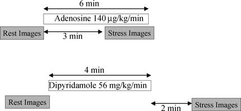
2 min after completion of infusion. The same infusion line Because ischaemia-induced wall motion abnormalities may is used to administer the contrast agent and the vasodilator.
resolve quickly, post-treadmill exercise imaging should be Alternatively, an infusion of adenosine 140 mg/kg/min (half- accomplished within 60–90 s of termination of exercise.
life 4–10 s) can be used. The contrast echocardiograms are Therefore, it is necessary to inject the bolus (if only wall acquired prior to infusion and again during the infusion at motion is assessed) or start the infusion (ideal for assessing 3 min. Usually, all the three apical views + available para- perfusion) of contrast before the patient terminates the sternal views can be acquired within the subsequent exercise. After application of the contrast agent, the 3 min, resulting in a total adenosine infusion time of 6 min patient should be asked to continue the exercise for at (Figure 7). For both vasodilators, a three-way tap or small least 20 s, before laying down for image acquisition.
bore Y connector is useful to connect the vasodilator infu-sion lines and contrast infusion pump.
Dobutamine stress echocardiography The protocol for native dobutamine stress echocardiography Myocardial contrast echocardiography is described elsewhere except apical views are acquired first for myocardial viability (Figure 6).89 Resting image settings should be optimized and Rest contrast images are acquired as described previously.
resting contrast echocardiogram views should be obtained However, when real-time imaging protocol is used, it is according to the criteria described previously. The same important to acquire at least 15 sc cycle post-flash for views are acquired at an intermediate stage (70% of the optimal assessment. It has been shown that both the pre- maximum age predicted heart rate) and at peak stress (85% sence of homogenous contrast uptake and, alternatively, of the maximum age predicted heart rate). Contrast can be the absence of contrast uptake are very accurate indicators administered as a bolus or as an infusion, as described pre- of the presence or absence of myocardial viability, respect- viously. If the SonoVue-infusion pump or Luminity drip is ively. It is, however, important to exclude apical and basal used, infusion is needed only while acquiring the images.
artefacts before concluding that there is absent contrast Prior to acquisition of the peak stress loops, the contrast infu- uptake. High MI imaging should also include imaging up to sion is started and the dobutamine infusion is stopped. Within 15 s intermittent image acquisition. The transmit focus 30 s, there is sufficient contrast enhancement and the peak should be moved towards the apex to confirm an apical per- contrast images are acquired in the apical views (and para- fusion defect when suspected. A thin and scarred (bright) sternal views if of sufficient quality). Image acquisition at myocardium of ,5 mm in size indicates non-viable tissue stress is not different from acquisition at rest except when and it is unnecessary to assess perfusion in these segments.
using triggered imaging. A 1:1 trigger interval should beused at peak stress whereas at rest 1:4 is useful. Adjustment of the time delay of the trigger may be necessary at peakstress to allow for the increased heart rate.
Lefrt ventricular function and regional wall motionVisual analysis of LV function is performed in all patientsaccording Vasodilator stress Vasodilator stress is the best stress modality enhancement of the epicardial vessels helps in judging for perfusion imaging wall thickening as well as inward motion of the myocardium.
An amount of 0.56 mg/kg of dipyridamole (half-life 30 min) Contrast images are ideal for measuring LV volumes and LVEF. The post-flash (destruction) images provide the best R. Senior et al.
Protocol for myocardial contrast echocardiography. Protocol 1.
contrast between myocardium and the LV cavity. Manual Myocardial perfusion tracing on still frames to obtain LV volumes and ejection Although there is growing evidence of the usefulness of fraction is easy and quick. Therefore, in every contrast quantitative analysis,92 myocardial contrast signals are cur- study, these measurements should be obtained. The tools rently judged using visual assessment.
for automatic assessment of LV borders (such as colour Normal myocardial perfusion is displayed by homogeneous kinesis), as well as 3D echocardiographic volumetric assess- contrast enhancement at rest 5 s after flash (Low MI ments with contrast are currently being investigated in clini- imaging) or high MI imaging and a quick replenishment at cal trials and may be useful clinical tools in the future.90,91 stress (within 2 s).


Evidence-based recommendations by EAE on contrast echocardiography Protocol for myocardial contrast echocardiography. Protocol 2.
have the same or worse imaging conditions. The diagnosticconfidence of an observed perfusion defect increases when Dobutamine stress contrast protocol.
two contiguous segments fail to exhibit contrast enhance-ment. A contrast defect is usually seen first in the subendo- Perfusion defect by myocardial contrast cardium and does not extend over the full thickness of the myocardium. The specificity for the detection of a perfusion A visually evident contrast defect is considered present defect is decreased in the absence of at least some contrast when there is a relative decrease in contrast enhancement signal in the epicardium; full thickness defects in which both in one region compared with other adjacent regions that endocardium and epicardium are absent are more likely to R. Senior et al.
be due to artefact such as attenuation or rib shadowing.
intervention. Combined assessment of wall motion and per- Basal lateral and anterior walls quite often cannot be fusion can also help to increase the diagnostic confidence of assessed because of these limitations. However, there is usually enough adequate information in other segments accompanied by a perfusion defect suggests flow-limiting in the LAD or RCX perfusion territories to permit the CAD. A new wall motion abnormality without a correspond- assessment of adequacy of perfusion by coronary artery ing perfusion defect may suggest cardiomyopathy.
Training and accreditation Fixed vs. reversible perfusion defect Both physicians and cardiac sonographers must have Fixed perfusion defects are visible at rest and stress. Revers- acquired basic echocardiography training and preferably ible defects are best seen early after the flash (during the accredited in echocardiography before using contrast first 2 cardiac cycles using real-time imaging) or with low agents. Those planning to use contrast agents during stress trigger rate (1:1 using high power triggered imaging).
echocardiography must be accredited or at least must Reversible defects suggestive of CAD are characterized by have undergone equivalent training in stress echocardiogra- delayed subendocardial replenishment and subendocardial phy. Beyond these trainings in rest and stress echocardiogra- hypoenhancement. With longer replenishment time (low MI phy, the use of contrast agents requires a level of experience imaging) or increased trigger intervals (high MI imaging), and performance, initially under guidance or supervision.
reversible defects often decrease in size or fill in.
Physicians, sonographers, and nurses alike should be compe- When assessing for potential perfusion defects, it is tent in the administration of contrast agents, should be crucial to avoid either oversaturation with contrast, or aware of the indications and contraindications, and should alternatively, inadequate concentration of contrast. Subtle be able to manage adverse events. It is encouraged that per- subendocardial defects may be obscured by excess contrast.
sonnel involved in contrast use should attend courses, etc.
These may be revealed by a reduction in contrast infusion to learn and familiarize themselves with the use of contrast, rate or further bubble destruction with additional intermit- performance, and interpretation of contrast-enhanced tent high power frames. On the other hand, inadequate con- images. This is particularly important if the echo team is trast concentration will obviate the detection of normally contemplating using contrast for assessment of perfusion perfused and relatively underperfused regions.
also.127,128 The echo team should seek guidance from thelocal echocardiography society of institutional director todetermine ways to set up myocardial perfusion programme.
Integrating wall motion findings and judgment It cannot be emphasized more that experience with contrast agent for LVO is a prerequisite for moving on to assess per- During rest echocardiography (Table 10) fusion and function with contrast agents.
This may be particularly useful when the assessment of LVwall motion is difficult or dubious at rest. Probably, the most important situation where perfusion imaging makes adifference is in akinetic areas (Table 7). Before one tries Contrast echocardiography significantly improves the image to assess myocardial contrast enhancement, it is important quality during rest and stress echocardiography and at the to look at myocardial thickness that often can be seen same time provides additional information on myocardial very well during infusion of contrast. When analysing loops perfusion. Contrast echocardiography reduces the need for obtained with real-time imaging, judgement of wall additional, costly, and more hazardous tests and, impor- motion and myocardial perfusion is often combined. A sub- tantly, spares the patient further invasive investigations.
endocardial perfusion defect makes a wall motion abnormal- Thus, contrast echocardiography provides a safe and com- ity much clearer and vice versa. Thus, assessment of prehensive assessment of cardiac structure, function, per- myocardial contrast often helps by increasing the diagnostic fusion, and coronary flow reserve at the bedside.
confidence of dubious wall motion analysis. In a restingstudy, a perfusion defect can be due to ischaemia with aflow limiting coronary stenosis at rest, a scar, or an artefact.
Conflict of interest: R.S. received research support from Bracco, Artefacts are most likely to occur in the basal lateral and Acusphere. M.M. has received research support from Bracco, Acu- anterior walls and can easily be detected by the typical cri- sphere, Philips, GE, Siemens; Speaker's Bureau: Philips, Siemens.
teria of attenuation and shadowing, in the presence P.N. has received research grants from Bracco and Medtronic. H.B.
acts as a consultant for Lantheaus, Bracco, POINT, Acusphere and of normal myocardial wall thickening. Other discrepant has research grants from Phillips, Sonosite, Siemens, Toshiba.
findings between wall motion and myocardial contrastenhancement occur with stunning and hibernation. Both con-ditions can be suspected when reduced wall motion and good contrast enhancement are found in a resting perfusion study.
1. Olszewski R, Timperley J, Cezary S, Monaghan M, Nihoyannopoulos P, Senior R et al. The clinical applications of contrast echocardiography.
During stress echocardiography (Table 11) Eur J Echocardiogr 2007;8:S13–S23.
Concordant findings in wall motion and perfusion increase 2. Mulvagh SL, Rakowski H, Vannan MA, Abdelmoneim SS, Becher H, our confidence when assessing a dubious wall motion Bierig M et al. ASE consensus statement on the clinical applications of abnormality. For instance, when asked for the significance ultrasonic contrast agents in echocardiography. J Am Soc Echocardiog2008;21:1179–1201.
of an angiographically determined coronary stenosis, 3. Thanigaraj S, Nease RF, Schechtman KB et al. Use of contrast for image normal wall motion and perfusion in the territory supplied enhancement during stress echocardiography is cost-effective and by this vessel should negate the need for coronary reduces additional diagnostic testing. Am J Cardiol 2001;87:1430–2.
Evidence-based recommendations by EAE on contrast echocardiography 4. Main ML, Grayburn PA. Clinical applications of transpulmonary contrast assessment in 300 consecutive patients. J Am Soc Echocardiogr 2001; echocardiography. Am Heart J 1999;137:144–53.
5. McCulloch M, Gresser C, Moos S, Odabashian J, Jasper S, Bednarz J et al.
26. Moir S, Haluska BA, Jenkins C, Fathi R, Marwick TH. Incremental benefit Ultrasound contrast physics: a series on contrast echocardiography.
of myocardial contrast to combined dipyridamole-exercise stress echo- J Am Soc Echocardiogr 2000;13:959–67.
cardiography for the assessment of coronary artery disease. Circulation 6. EMEA Scientific Discussion. SonoVue, European Public Assessment 27. Calachanis M, Evdoridis C, Nihoyannopoulos P. Improved diagnostic AR/SonoVue/SonoVue.htm (Accessed on 17 September).
accuracy of dobutamine stress echocardiography with Echogen.
7. GE Healthcare. OPTISON Prescribing Information. 2008; http://www.
Cardiovasc Imag 1999;11:123.
amershamhealth-us.com/optison/ (Accessed on 17 September).
28. Plana JC, Mikati IA, Dokainish H, Lakkis N, Abukhalil J, Davis R et al.
8. Luminity Summary of Product Characteristics. 2008 A randomized cross-over study for evaluation of the effect of image 9. Senior R, Kaul S, Soman P, Lahiri A. Power doppler harmonic imaging: a optimization with contrast on the diagnostic accuracy of dobutamine feasibility study of a new technique for the assessment of myocardial echocardiography in coronary artery disease. J Am Coll Cardiol perfusion. Am Heart J 2000;139:245–51.
Cardiac Imaging 2008;1:145–52.
10. Kuersten B, Murthy TH, Li P, Liu Z, Locricchio E, Baisch C et al. Ultrahar- 29. Jeetley P, Burden L, Stoykova B, Senior R. Clinical and economic impact monic myocardial contrast imaging: in vivo experimental and clinical of stress echocardiography compared to exercise electrocardiography in data from a novel technique. J Am Soc Echocardiogr 2001;14:910–6.
patients with suspected acute coronary syndrome but negative tropo- 11. Sieswerda GT, Yang L, Boo MB et al. Real-time perfusion imaging: a new nin: a prospective randomised controlled study. Eur Heart J 2007;28: echocardiographic technique for simultaneous evaluation of myocardial perfusion and contraction. Echocardiography 2003;20:545–55.
30. Thanigaraj S, Nease RF Jr, Schechtman KB, Wade RL, Loslo S, Pe 12. Bristol-Myers Squibb. Data on file, Definity Common Technical Docu- Use of contrast for image enhancement during stress echocardiography ment. Module 2: 2.7.3 Summary of Clinical Efficacy.
is cost-effective and reduces additional diagnostic testing. Am J Cardiol 13. Hundley WG, Kizilbash AM, Afridi I, Franco F, Peshock RM, Grayburn PA.
Administration of an intravenous perfluorocarbon contrast agent 31. Vlassak I, Rubin DN, Odabashian JA, Garcia MJ, King LM, Lin SS et al. Con- improves echocardiographic determination of left ventricular volumes trast and harmonic imaging improves accuracy and efficiency of novice and ejection fraction: comparison with cine magnetic resonance readers for dobutamine stress echocardiography. Echocardiography imaging. J Am Coll Cardiol 1998;32:1426–32.
14. Reilly JP, Tunick PA, Timmermans RJ, Stein B, Rosenzweig BP, Kronzon I.
32. Soman P, Swinburn J, Callister M, Stephens NG, Senior R. Apical hyper- Contrast echocardiography clarifies uninterpretable wall motion in trophic Cardiomyopathy: bedside diagnosis by intravenous contrast intensive care unit patients. J Am Coll Cardiol 2000;35:485–90.
echocardiography. J Am Soc Echocardiogr 2001;14:311–3.
15. Nahar T, Croft L, Shapiro R, Fruchtman S, Diamond J, Henzlova M et al.
33. Thanigaraj S, Schechtman KB, Perez JE. Improved echocardiographic Comparison of four echocardiographic techniques for measuring left delineation of left ventricular thrombus with the use of intravenous ventricular ejection fraction. Am J Cardiol 2000;86:1358–62.
second-generation contrast image enhancement. J Am Soc Echocardiogr 16. Yu EH, Sloggett CE, Iwanochko RM, Rakowski H, Siu SC. Feasibility and accuracy of left ventricular volumes and ejection fraction determi- 34. Koo BK, Choi D, Ha JW, Kang SM, Chung N, Cho SY. Isolated noncompac- nation by fundamental, tissue harmonic, and intravenous contrast tion of the ventricular myocardium: contrast echocardiographic findings imaging in difficult-to-image patients. J Am Soc Echocardiogr 2000; and review of the literature. Echocardiography 2002;19:153–6.
35. Mittle S, Makaryus AN, Mangion J. Role of contrast echocardiography in 17. Yong Y, Wu D, Fernandes V, Kopelen HA, Shimoni S, Nagueh SF et al.
the assessment of myocardial rupture. Echocardiography 2003;20: Diagnostic accuracy and cost-effectiveness of contrast echocardiogra- phy on evaluation of cardiac function in technically very difficult 36. Kusnetzky LL, Khalid A, Khumri TM, Moe TG, Jones PG, Main ML. Acute patients in the intensive care unit. Am J Cardiol 2002;89:711–8.
mortality in hospitalized patients undergoing echocardiography with 18. Malm S, Frigstad S, Sagberg E, Larsson H, Skjaerpe T. Accurate and and without an ultrasound contrast agent. J Am Coll Cardiol 2008;51: reproducible measurement of left ventricular volume and ejection frac- tion by contrast echocardiography: a comparison with magnetic reson- 37. Timperley J, Mitchell AR, Thibault H, Mirza IH, Becher H. Safety of con- ance imaging. J Am Coll Cardiol 2004;44:1030–5.
trast dobutamine stress echocardiography: a single center experience.
19. Hoffmann R, von Bardeleben S, ten Cate F, Borges AC, Kasprzak J, J Am Soc Echocardiogr 2005;18:163–7.
Firschke C et al. Assessment of systolic left ventricular function: a 38. Anantharam B, Chahal N, Chelliah R, Ramzy I, Gani F, Senior R. Safety of multi-centre comparison of cineventriculography, cardiac magnetic res-onance imaging, unenhanced and contrast-enhanced echocardiography.
contrast in stress echocardiography in stable patients and in patients Eur Heart J 2005;26:607–16.
with suspected acute coronary syndrome but negative 12 hour Troponin.
20. Lim TK, Burden L, Janardhanan R, Ping C, Moon J, Pennell D et al.
Am J Cardiol 2009 (in press).
Improved accuracy of low-power contrast echocardiography for the 39. Tsutsui JM, Elhendy A, Xie F, O'Leary EL, McGrain AC, Porter TR. Safety assessment of left ventricular remodeling compared with unenhanced of dobutamine stress real-time myocardial contrast echocardiography.
harmonic echocardiography after acute myocardial infarction: compari- J Am Coll Cardiol 2005;19:1235–42.
son with cardiovascular magnetic resonance imaging. J Am Soc 40. Aggeli C, Giannopoulos G, Roussakis G, Christoforatou E, Marinos G, Toli C et al. Safety of myocardial flash-contrast echocardiography in 21. Weiss RJ, Lieux TR, Ahmad M, Shirani J. An open-label, randomised, combination with dobutamine stress testing for detection of ischemia multi-centre trial to examine the predictive value of Definity contrast in 5250 studies. Heart 2008;94:1571–7.
stress echocardiography on patient outcomes. J Am Soc Echocardiogr 41. Main ML, Goldman JH, Grayburn PA. Thinking outside the ‘box'—the ultrasound contrast controversy. J Am Coll Cardiol 2007;18:2434–7.
22. Shaw LJ, Monaghan MJ, Nihoyannopoulos P. Clinical and economic out- 42. Wei K, Mulvagh SL, Carson L, Davidoff R, Gabriel R, Grim RA et al. The comes assessment with myocardial contrast echocardiography. Heart safety of Definity and Optison for ultrasound image enhancement: a ret- rospective analysis of 78,383 administered contrast doses. J Am Soc 23. Dolan MS, Riad K, El-Shafei A, Puri S, Tamirisa K, Bierig M et al. Effect of intravenous contrast for left ventricular opacification and border defi- 43. Kaul S, Jayaweera AR. Coronary and myocardial blood volumes: nonin- nition on sensitivity and specificity of dobutamine stress echocardiogra- vasive tools to assess the coronary microcirculation. Circulation 1997; phy compared with coronary angiography in technically difficult patients. Am Heart J 2001;142:908–15.
44. Wei K, Jayaweera AR, Firoozan S, Linka A, Skyba DM, Kaul S. Basis for 24. Shimoni S, Zoghbi WA, Xie F, Kricsfeld D, Iskander S, Gobar L et al. Real- detection of stenosis using venous administration of microbubbles time assessment of myocardial perfusion and wall motion during bicycle during myocardial contrast echocardiography: bolus or continuous infu- and treadmill exercise echocardiography: comparison with single sion? J Am Coll Cardiol 1998;32:252–60.
photon emission computed tomography. J Am Coll Cardiol 2001;37: 45. Wei K, Jayaweera AR, Firoozan S, Linka A, Skyba DM, Kaul S. Quantifi- cation of myocardial blood flow with ultrasound-induced destruction 25. Rainbird AJ, Mulvagh S, Oh JK, McCully RB, Klarich KW, Shub C et al.
of microbubbles administered as a constant venous infusion. Circulation R. Senior et al.
46. Kaul S, Senior R, Dittrich H, Raval U, Khattar R, Lahiri A. Detection of with chest pain and a nondiagnostic electrocardiogram. J Am Coll coronary artery disease with myocardial contrast echocardiography: comparison with 99mTc-sestamibi single-photon emission computed tom- 64. Rinkevich D, Kaul S, Wang X-Q et al. Regional left ventricular perfusion ography. Circulation 1997;96:785–92.
and function in patients presenting to the emergency department with 47. Heinle SK, Noblin J, Goree-Best P, Mello A, Ravad G, Mull S et al. Assess- chest pain and no ST-segment elevation. Eur Heart J 2005;26:1606–11.
ment of myocardial perfusion by harmonic power Doppler imaging at 65. Jeetley P, Burden L, Greaves K, Senior R. Prognostic value of myocardial rest and during adenosine stress: comparison with 99mTc-sestamibi contrast echocardiography in patients presenting to hospital with acute SPECT imaging. Circulation 2000;102:55–60.
chest pain and negative troponin. Am J Cardiol 2007;99:1369–73.
48. Shimoni S, Zoghbi WA, Xie F, Kricsfeld D, Iskander S, Gobar L et al. Real- 66. Ragosta M, Camarano GP, Kaul S, Powers E, Sarembock IJ, Gimple LW.
time assessment of myocardial perfusion and wall motion during bicycle Microvascular integrity indicates myocellular viability in patients with and treadmill exercise echocardiography: comparison with single recent myocardial infarction: new insights using myocardial contrast photon emission computed tomography. J Am Coll Cardiol 2001;37: 67. Shimoni S, Frangogiannis NG, Aggeli CJ et al. Microvascular structural 49. Wei K, Crouse L, Weiss J, Villanueva F, Schiller NB, Naqvi TZ et al. Com- correlates of myocardial contrast echocardiography in patients with cor- parison of usefulness of dipyridamole stress myocardial contrast echo- onary artery disease and left ventricular dysfunction: implications for cardiography to technetium-99m sestamibi single-photon emission the assessment of myocardial hibernation. Circulation 2002;108:950–6.
computed tomography for detection of coronary artery disease (PB127 68. Coggins MP, Sklenar J, Le DE, Wei K, Lindner JR, Kaul S. Noninvasive pre- Multicenter Phase 2 Trial results). Am J Cardiol 2003;91:1293–8.
diction of ultimate infarct size at the time of acute coronary occlusion 50. Rocchi G, Fallani F, Bracchetti G, Rapezzi C, Ferlito M, Levorato M et al.
based on the extent and magnitude of collateral-derived myocardial Non-invasive detection of coronary artery stenosis: a comparison among blood flow. Circulation 2001;104:2471–7.
power-Doppler contrast echo, 99Tc-Sestamibi SPECT and echo wall- 69. Lafitte S, Higashiyama A, Masugata H, Peters B, Strachan M, Kwan OL motion analysis. Coron Artery Dis 2003;14:239–45.
et al. Contrast echocardiography can assess risk area and infarct size 51. Olszowska M, Kostkiewicz M, Tracz W, Przewlocki T. Assessment of myo- during coronary occlusion and reperfusion: experimental validation.
cardial perfusion in patients with coronary artery disease. Comparison J Am Coll Cardiol 2002;39:1546–54.
of myocardial contrast echocardiography and 99mTc MIBI single photon 70. Janardhanan R, Moon JC, Pennell DJ, Senior R. Myocardial contrast emission computed tomography. Int J Cardiol 2003;90:49–55.
echocardiography accurately reflects transmurality of myocardial 52. Senior R, Lepper W, Pasquet A, Chung G, Hoffman R, Vanoverschelde JL necrosis and predicts contractile reserve after acute myocardial infarc- et al. Myocardial perfusion assessment in patients with medium prob- tion. Am Heart J 2005;149:355–62.
ability of coronary artery disease and no prior myocardial infarction: 71. Choi EY, Seo HS, Park S et al. Prediction of transmural extent of infarc- comparison of myocardial contrast echocardiography with 99mTc single- tion with contrast echocardiographically derived index of myocardial photon emission computed tomography. Am Heart J 2004;147:1100–5.
blood flow and myocardial blood volume fraction: comparison with 53. Xie F, Tsutsui JM, McGrain AC, DeMaria A, Cotter B, Becher H et al. Com- contrast-enhanced magnetic resonance imaging. J Am Soc Echocardiogr parison of dobutamine stress echocardiography with and without real- time perfusion imaging for detection of coronary artery disease. Am J 72. Hickman M, Burden L, Senior R. Resting myocardial blood flow not myo- cardial blood flow reserve predicts hibernating myocardium: A quanti- 54. Korosoglou G, Dubart AE, DaSilva KG Jr, Labadze N, Hardt S, Hansen A tative myocardial contrast echocardiography study. J Am Coll Cardiol et al. Real-time myocardial perfusion imaging for pharmacologic stress testing: added value to single photon emission computed 73. Senior R, Swinburn JM. Incremental value of myocardial contrast echo- tomography. Am Heart J 2006;151:131–8.
cardiography for the prediction of recovery of function in dobutamine 55. Dijkmans PA, Senior R, Becher H, Porter TR, Wei K, Visser CA et al. Myo- nonresponsive myocardium early after acute myocardial infarction.
cardial contrast echocardiography evolving as a clinically feasible tech- Am J Cardiol 2003;91:397–402.
nique for accurate, rapid, and safe assessment of myocardial perfusion: 74. Shimoni S, Frangogiannis NG, Aggeli CJ et al. Identification of hibernat- the evidence so far. J Am Coll Cardiol 2006;48:2168–77.
ing myocardium with quantitative intravenous myocardial contrast 56. Senior R, Monaghan M, Main ML, Zamorano JL, Tiemann K, Agali L et al.
echocardiography: comparison with dobutamine echocardiography and Detection of coronary artery disease with perfusion stress echocardio- thallium-201 scintigraphy. Circulation 2003;107:538–44.
graphy using a novel ultrasound imaging agent: two phase 3 inter- 75. Greaves K, Dixon SR, Fejka M, O'Neill WW, Redwood SR, Marber MS et al.
national trials in comparison with radionuclide perfusion imaging. Eur Myocardial contrast echocardiography is superior to other known modal- J Echocardiogr 2009;10:26–35.
ities for assessing myocardial reperfusion after acute myocardial infarc- 57. Marwick TH, Brunken R, Meland N, Brochet E, Baer FM, Binder T et al.
tion. Heart 2003;89:139–44.
Accuracy and feasibility of contrast echocardiography for detection of 76. Bolognese L, Carrabba N, Parodi G et al. Impact of microvascular dys- perfusion defects in routine practice: Comparison with wall motion function on left ventricular remodeling and long-term clinical and Technitium-99, Sestamibi Single Photon Emission Computed Tom- outcome after primary coronary angioplasty for acute myocardial ography. J Am Coll Cardiol 1998;32:1260–9.
infarction. Circulation 2004;109:1121–6.
58. Tsutsui JM, Elhendy A, Anderson JR, Xie F, McGrain AC, Porter TR. Prog- 77. Galiuto L, Garramone B, Scara A, Rebuzzi A, Crea F, La Torre G et al. The nostic value of dobutamine stress myocardial contrast perfusion echo- extent of microvascular damage during myocardial contrast echocardio- graphy is superior to other known indexes of post-infarct reperfusion in 59. Sabia P, Abbott RD, Afrookteh A, Keller MW, Touchstone DA, Kaul S.
predicting left ventricular remodeling: results of the multicenter AMICI Importance of two-dimensional echocardiographic assessment of left study. J Am Coll Cardiol 2008;51:552.
ventricular systolic function in patients presenting to the emergency 78. Ito H, Maruyama A, Iwakura K, Takiuchi S, Masuyama T, Hori M et al.
room with cardiac-related symptoms. Circulation 1991;84:1615–24.
Clinical implications of the ‘no reflow' phenomenon. A predictor of 60. Kaul S, Senior R, Firschke C, Wang XQ, Lindner J, Villanueva FS et al.
complications and left ventricular remodeling in reperfused anterior Incremental value of cardiac imaging in patients presenting to the wall myocardial infarction. Circulation 1996;93:223–8.
emergency department with chest pain and without ST-segment 79. Khumri T, Nayyar S, Idupulapati M, Magalski A, Stoner C, Kusnetzky L elevation: a multicenter study. Am Heart J 2004;148:129–36.
et al. Usefulness of Myocardial Contrast Echocardiography in Predicting 61. Kontos MC, Hinchman D, Cunningham M, Miller JJ, Cherif J, Nixon JV.
Late Mortality in Patients With Anterior Wall Acute Myocardial Infarc- Comparison of contrast echocardiography with single-photon emission tion. Am J Cardiol 2006;98:1150–5.
computed tomographic myocardial perfusion imaging in the evaluation 80. Dwivedi G, Janardhanan R, Hayat S, Swinburn J M, Senior R. Prognostic of patients with possible acute coronary syndromes in the emergency value of myocardial viability detected by myocardial contrast echocar- department. Am J Cardiol 2003;91:1099–102.
diography early after acute myocardial infarction. J Am Coll Cardiol 62. Tsutsui JM, Xie F, O'Leary EL, Elhendy A, Anderson JR, McGrain AC et al.
Diagnostic accuracy and prognostic value of dobutamine stress myocar- 81. Hayat SA, Senior R. Myocardial contrast echocardiography in ST dial contrast echocardiography in patients with suspected acute coron- elevation myocardial infarction: ready for prime time? Eur Heart J ary syndromes. Echocardiography 2005;22:487–95.
63. Tong KL, Kaul S, Wang XQ, Rinkevich D, Kalvaitis S, Belcik T et al. Myo- 82. Wei K, Ragosta M, Thorpe J, Coggins M, Moos S, Kaul S. Non-invasive cardial contrast echocardiography versus Thrombolysis In Myocardial quantification of coronary blood flow reserve in humans using myocar- Infarction score in patients presenting to the emergency department dial contrast echocardiography. Circulation 2001;103:2560–5.
Evidence-based recommendations by EAE on contrast echocardiography 83. Peltier M, Vancraeynest D, Pasquet A, Ay T, Roelants V, D'hondt AM et al.
stress testing: added value to single photon emission computed Assessment of the physiologic significance of coronary disease with tomography. Am Heart J 2006;151:131–8.
dipyridamole real-time myocardial contrast echocardiography. Compari- son with technetium-99m sestamibi single-photon emission computed Nihoyannopoulos P. Left ventricular wall motion assessment and Endo- tomography and quantitative coronary angiography. J Am Coll Cardiol cardial border delineation after intravenous injection of InfosonTM during dobutamine stress echocardiography. Coronary Artery Disease 84. Vogel R, Indermuhle A, Reinhardt J, Meier P, Siegrist PT, Namdar M et al.
The quantification of absolute myocardial perfusion in humans by con- 104. Bhatia V, Senior R. Contrast echocardiography in clinical practice. Evi- trast echocardiography. J Am Coll Cardiol 2005;45:754–62.
dence so far. J Am Soc Echocardiogr 2008;21:409–16.
85. Senior R, Janardhanan R, Jeetley P, Burden L. Myocardial contrast echo- 105. Chiou KR, Huang WC, Lin SL, Hsieh PL, Liu CP, Tsay DG et al. Real-time cardiography for distinguishing ischemic from nonischemic first-onset dobutamine stress myocardial contrast echocardiography for detecting acute heart failure: insights into the mechanism of acute heart coronary artery disease: correlating abnormal wall motion and dis- failure. Circulation 2005;112:1587–93.
turbed perfusion. Can J Cardiol 2004;20:1237–43.
86. Hayat SA, Dwivedi G, Jacobsen A, Kinsey C, Senior R. Effects of left 106. Cwajg J, Xie F, O'Leary E, Kricsfeld D, Dittrich H, Porter TR. Detection of bundle branch block on cardiac structure, function perfusion and per- angiographically significant coronary artery disease with accelerated fusion reserve: implications for myocardial contrast echocardiography intermittent imaging after intravenous administration of ultrasound versus radionuclide perfusion imaging for the detection of coronary contrast material. Am Heart J 2000;139:675–83.
artery disease. Circulation 2008;117:1832–41.
107. Elhendy A, O'Leary EL, Xie F, McGrain AC, Anderson JR, Porter TR. Com- 87. Janardhanan R, Senior R. Accuracy of dipyridamole myocardial contrast parative accuracy of real-time myocardial contrast perfusion imaging echocardiography for the detection of residual stenosis of the and wall motion analysis during dobutamine stress echocardiography infarct-related artery and multivessel disease early after acute myocar- for the diagnosis of coronary artery disease. J Am Coll Cardiol 2004; dial infarction. J Am Coll Cardiol 2004;43:2247–52.
88. Moir S, Haluska BA, Jenkins C, McNab D, Marwick TH. Myocardial blood 108. Jeetley P, Hickman M, Kamp O, Lang RM, Thomas JD, Vannan MA et al.
volume and perfusion reserve responses to combined dipyridamole and Myocardial contrast echocardiography for the detection of coronary exercise stress: a quantitative approach to contrast stress echocardio- artery stenosis: a prospective multicenter study in comparison with graphy. J Am Soc Echocardiogr 2005;18:1187–93.
single-photon emission computed tomography. J Am Coll Cardiol 2006; 89. Sicari R, Nihoyannopoulos P, Evangelista A, Kasprzak J, Lancellotti P, Poldermans D et al. Stress echocardiography expert consensus state- 109. Karavidas AI, Matsakas EP, Lazaros GA, Brestas PS, Avramidis DA, ment: European Association of Echocardiograph (EAE) (a registered Zacharoulis AA et al. Comparison of myocardial contrast echocardiogra- branch of the ESC). Eur J Echocardiogr 2008;9:415–37.
phy with SPECT in the evaluation of coronary artery disease in asympto- 90. Senior R, Dwivedi G, Hayat S, Lim TH. Clinical benefits of matic patients with LBBB. Int J Cardiol 2006;112:334–40.
contrast-enhanced echocardiography during rest and stress examin- 110. Lin SL, Chiou KR, Huang WC, Peng NJ, Tsay DG, Liu CP. Detection of cor- ations. Eur J Echocardiogr 2005;6:S6–S13.
onary artery disease using real-time myocardial contrast echocardiogra- phy: a comparison with dual-isotope resting thallium-201/stress Nihoyannopoulos P, Senior R et al. The clinical applications of contrast technetium-99m sestamibi single-photon emission computed tomogra- echocardiography. Eur J Echocardiogr 2007;8:S13–S23.
phy. Heart Vessels 2006;21:226–35.
92. Rakhit DJ, Becher H, Monaghan M, Nihoyannopoulis P, Senior R. The 111. Malm S, Frigstad S, Torp H, Wiseth R, Skjarpe T. Quantitative adenosine clinical applications of myocardial contrast echocardiography. Eur real-time myocardial contrast echocardiography for detection of angio- J Echocardiography 2007;8:524–9.
graphically significant coronary artery disease. J Am Soc Echocardiogr 93. Tsutsui JM, Xie F, McGrain, Mahrous H, Hankins J, O'Leary EL et al. Com- parison of low-mechanical index pulse sequence schemes for detecting 112. Winter R, Gudmundsson P, Willenheimer R. Real-time perfusion adeno- myocardial perfusion abnormalities during vasodilator stress echocar- sine stress echocardiography in the coronary care unit: a feasible diography. Am J Cardiol 2005;95:565–70.
bedside tool for predicting coronary artery stenosis in patients with 94. Cohen JL, Cheirif J, Segar DS, Gillam LD, Gottdiener JS, Hausnerova E acute coronary syndrome. Eur J Echocardiogr 2005;6:31–40.
et al. Improved left ventricular endocardial border delineation and 113. Sbano JC, Tsutsui JM, Andrade JL, Carlos Nicolau J, Meneghetti JC, Fran- opacification with Optison (FS069), a new echocardiographic contrast chini Ramires J et al. Detection of functional recovery using low-dose agent. Results of a phase III multicenter trial. J Am Coll Cardiol 1998; dobutamine and myocardial contrast echocardiography after acute myocardial infarction treated with successful thrombolytic therapy.
95. Senior R, Andersson O, Caidahl K, Carlens P, Herregods MC, Jenni R et al.
Enhanced left ventricular endocardial border delineation with an intra- 114. Aggeli C, Stefanadis C, Bonou M, Pitsavos C, Theocharis C, Roussakis G venous injection of SonoVue, a new echocardiographic contrast agent: a et al. Prediction of functional recovery of hibernating myocardium European multicenter study. Echocardiography 2000;17:705–11.
using harmonic power Doppler imaging and dobutamine stress echocar- 96. Kitzman DW, Goldman ME, Gillam LD, Cohen JL, Aurigemma GP, diography in patients with coronary artery disease. Am J Cardiol 2003; Gottdiener JS. Efficacy and safety of the novel ultrasound contrast agent perflutren (Definity) in patients with suboptimal baseline left ven- 115. Hillis GS, Mulvagh SL, Pellikka PA, Hagen ME, Gunda M, Wright RS et al.
tricular echocardiographic images. Am J Cardiol 2000;86:669–74.
Comparison of intravenous myocardial contrast echocardiography and 97. Nguyen TT, Dhond MR, Sabapathy R, Bommer WJ. Contrast microbubbles low-dose dobutamine echocardiography for predicting left ventricular improve diagnostic yield in ICU patients with poor echocardiographic windows. Chest 2001;120:1287–92.
J Cardiol 2003;92:504–8.
98. Nanda NC, Wistran DC, Karlsberg RP, Hack TC, Smith WB, Foley DA et al.
116. Main ML, Magalski A, Chee NK, Coen MM, Skolnick DG, Good TH. Full- Multicenter evaluation of SonoVue for improved endocardial border motion pulse inversion power Doppler contrast echocardiography differ- entiates stunning from necrosis and predicts recovery of left ventricular 99. Rizzo M, Vono MC, Toncelli L, Pecagna P, Manetti P, Stefani L et al. The function after acute myocardial infarction. J Am Coll Cardiol 2001;38: feasibility and usefulness of contrast exercise echocardiography for the assessment of left ventricular function in master athletes. Eur J 117. Swinburn JM, Senior R. Real time contrast echocardiography—a new bedside technique to predict contractile reserve early after acute myo- 100. Tsutsui JM, Elhendy A, Xie F, O'Leary EL, McGrain AC, Porter TR. Safety cardial infarction. Eur J Echocardiogr 2002;3:95–9.
of dobutamine stress real-time myocardial contrast echocardiography.
118. Hillis GS, Mulvagh SL, Gunda M, Hagen ME, Reeder GS, Oh JK. Contrast J Am Coll Cardiol 2005;45:1235–42.
echocardiography using intravenous octafluoropropane and real-time 101. Wake R, Takeuchi M, Yoshitani H, Miyazaki C, Otani S, Yoshiyama M et al.
perfusion imaging predicts functional recovery after acute myocardial Role of contrast-enhanced dobutamine stress echocardiography in pre- infarction. J Am Soc Echocardiogr 2003;16:638–45.
dicting outcome in patients with known or suspected coronary artery 119. Lepper W, Kamp O, Vanoverschelde JL, Franke A, Sieswerda GT, disease. Echocardiography 2006;23:642–9.
Pasquet A et al. Intravenous myocardial contrast echocardiography pre- 102. Korosoglou G, Dubart AE, DaSilva KG, Labadze N, Hardt S, Hansen A dicts left ventricular remodeling in patients with acute myocardial et al. Real-time myocardial perfusion imaging for pharmacologic infarction. J Am Soc Echocardiogr 2002;15:849–56.
R. Senior et al.
120. Hickman M, Janardhanan R, Dwivedi G, Burden L, Senior R. Clinical sig- in predicting the left ventricular functional recovery in patients after nificance of perfusion techniques utilising different physiological mech- acute myocardial infarction under different therapeutic intervention.
anisms to detect myocardial viability: a comparative study with Int J Cardiol 2005;104:81–91.
myocardial contrast echocardiography and single photon emission com- 125. Abe Y, Muro T, Sakanoue Y et al. Intravenous myocardial contrast echo- puted tomography. Int J Cardiol 2007;114:139–40.
cardiography predicts regional and global left ventricular remodelling 121. Janardhanan R, Swinburn JM, Greaves K, Senior R. Usefulness of myo- after acute myocardial infarction: comparison with low dose dobuta- cardial contrast echocardiography using low-power continuous imaging mine stress echocardiography. Heart 2005;91:1578–83.
early after acute myocardial infarction to predict late functional left 126. Korosoglou G, Labadze N, Giannitsis E et al. Usefulness of real-time ventricular recovery. Am J Cardiol 2003;92:493–7.
myocardial perfusion imaging to evaluate tissue level reperfusion in 122. Main ML, Magalski A, Chee NK, Coen MM, Skolnick DG, Good TH. Full- patients with non-ST-elevation myocardial infarction. Am J Cardiol motion pulse inversion power Doppler contrast echocardiography differ- entiates stunning from necrosis and predicts recovery of left ventricular 127. Binder T, Assayag P, Baer F, Flachskampf F, Kamp O, Nienaber C et al.
function after acute myocardial infarction. J Am Coll Cardiol 2001;38: NC100100, a new echo contrast agent for the assessment of myocardial perfusion—safety and comparison with MIBI SPECT in a randomised mul- 123. Agati L, Voci P, Autore C et al. Combined use of dobutamine echocardio- ticentre study. Clin Cardiol 1999;22:273–82.
graphy and myocardial contrast echocardiography in predicting regional 128. Jucquois I, Nihoyannopoulos P, D'Hontdt A-M, Roelants V, Robert A, dysfunction recovery after coronary revascularization in patients with Melin JA et al. Comparison of myocardial contrast echocardiography recent myocardial infarction. Eur Heart J 1997;18:771–9.
with NC100100 and Tc-99m sestamibi single photon emission computed 124. Huang WC, Chiou KR, Liu CP et al. Comparison of real-time contrast tomography for detection of resting myocardial perfusion abnormalities echocardiography and low-dose dobutamine stress echocardiography in patients with previous myocardial infarction. Heart 2000;83:518–24.

Source: http://www.ceuscampus.de/images/stories/pdf-files/Evidenz_basierte_Empfehlungen_der_EAE_zur_Kontrast-Echokardiographie_2009.pdf

Effect of divalproex on brain morphometry, chemistry, and function in youth at high-risk for bipolar disorder: a pilot study

JOURNAL OF CHILD AND ADOLESCENT PSYCHOPHARMACOLOGYVolume 19, Number 1, 2009 ª Mary Ann Liebert, Inc.Pp. 51–59DOI: 10.1089=cap.2008.060 Effect of Divalproex on Brain Morphometry, Chemistry, and Function in Youth at High-Risk for Bipolar Disorder: A Pilot Study Kiki Chang, M.D., Asya Karchemskiy, M.S., Ryan Kelley, B.A., Meghan Howe, M.S.W., Amy Garrett, Ph.D., Nancy Adleman, B.S., and Allan Reiss, M.D.

trainorlab.ucdavis.edu

Author's personal copy Available online at www.sciencedirect.com Hormones and Behavior 53 (2008) 192 – 199 Rapid effects of estradiol on male aggression depend on photoperiod in reproductively non-responsive mice Brian C. Trainor a,b,⁎, M. Sima Finy b, Randy J. Nelson b a Department of Psychology, University of California, Davis, CA 95616, USA b Departments of Psychology and Neuroscience, Institute for Behavioral Medicine Research, Ohio State University, Columbus, OH 43210, USA