
Cialis ist bekannt für seine lange Wirkdauer von bis zu 36 Stunden. Dadurch unterscheidet es sich deutlich von Viagra. Viele Schweizer vergleichen daher Preise und schauen nach Angeboten unter dem Begriff cialis generika schweiz, da Generika erschwinglicher sind.
Doi:10.1016/j.mpsur.2007.10.013
The diabetic foot
infection can lead to tissue necrosis and amputation. Diabetes is the leading non-traumatic cause of major amputation of the lower limbs.
Miles J levyJonathan ValabhjiQ2
Neuropathy
Nerve damage due to disease of the vasa nervorum results in
a ‘glove and stocking' sensorimotor peripheral neuropathy that
may progress proximally. The motor component results in dener-
vation of the small muscles of the foot, leading to:
• hyperextension at the metatarsophalangeal joints
• hyperflexion at the interphalangeal joints
Foot disease is a common complication of type-1 and type-2 diabetes.
• cavus deformity of the arch of the foot.
The term ‘diabetic foot' refers to a spectrum of disease that includes the
These features produce the classical ‘claw foot' deformity
foot at risk of ulceration, the ulcerated foot, and the charcot foot. risk
. The neuropathic posture puts increased pres-
of ulceration is conferred by peripheral neuropathy and peripheral vas-
sure on the plantar aspects of the metatarsal heads, leading
cular disease. The ulcerated foot can be classified as neuropathic, neu-
to callus formation. Sensory neuropathy results in loss of the
roischaemic or ischaemic. The principles of ulcer management include
protective sensations of pain, heat and pressure, making the
diagnosis and treatment of infection, establishment and treatment of
patient unaware of actual or incipient ulceration. Autonomic
vascular insufficiency, off-loading and optimization of the wound envi-
neuropathy leads to vasomotor denervation and arteriovenous
ronment. The charcot foot is a rare complication of the neuropathic foot
shunting, compromising the ability to direct blood flow into
and off-loading in the acute phase is important to prevent destruction
capillary beds. The paradox of bounding peripheral pulses in
of joint architecture. long-established multidisciplinary foot clinics have
the presence of reduced microvascular flow may mask tissue
achieved up to 50% reductions in major amputations in the uK.
ischaemia. Autonomic denervation of sweat glands leads to dry skin and fissuring, which may provide a portal for microbial
Keywords charcot; diabetic foot; diabetic ulcer; neuroischaemic foot;
Ischaemia
Ischaemia can be due to large- or small-vessel disease.
Large-vessel disease is due to accelerated atherosclerosis,
usually in the femoral, popliteal and tibial arteries.
Foot disease is a common complication of Type 1 and Type 2
Small-vessel disease is due to structural and functional abnor-
diabetes mellitus. The term ‘diabetic foot' refers to a spectrum
malities of the microvascular endothelium. The skin of the foot
is red, dry and thin, and susceptible to breakdown on minimal
The foot at risk of ulceration – the risk of ulceration is con-
ferred by the diabetic microvascular complication of peripheral neuropathy, and the macrovascular complication of peripheral vascular disease.
The ulcerated foot can be neuropathic, neuroischaemic or
The Charcot foot is rare in the UK, but can lead to significant
bony destruction, deformity and ulceration.
About 20–40% of patients with diabetes have neuropathy, and 50% will develop symptomatic peripheral vascular disease within 20 years of diagnosis. The lifetime prevalence of foot ulceration in people with diabetes is about 15%. Ulceration with
Miles J Levy MBBS is a Consultant Physician, Department of
Endocrinology, Leicester Royal Infirmary, Leicester, UK. Conflicts of
interest: none declared.Q1
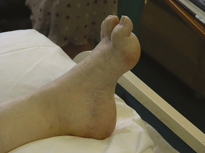
Figure 1 The classical ‘claw foot' deformity in diabetic neuropathy.
Jonathan Valabhji MBBS MRCP is a Consultant Physician, Department of
There is hyperextension at the metatarsophalangeal joint and
Endocrinology, St Mary's Hospital, Imperial College Healthcare NHS
hyperflexion at the interphalangeal joints. The arch of the foot has a
trust, London, UK. Conflicts of interest: none declared.
cavus deformity.
2007 Elsevier ltd. all rights reserved.
Contributory factors
In addition to neurovascular disease in the foot, a combination
of factors may contribute to the onset of ulceration in diabetic
patients, including:
• poor vision
• limited mobility in the joints
• cerebrovascular disease
• peripheral oedema due to coronary heart disease.
The foot at-risk
Clinical assessment
The diabetic foot should be assessed for neuropathy and peri-
pheral vascular disease.
Neuropathy is detected by:
• testing vibration with a biosthesiometer or tuning fork • discriminatory touch using a 10-g monofilament (highly
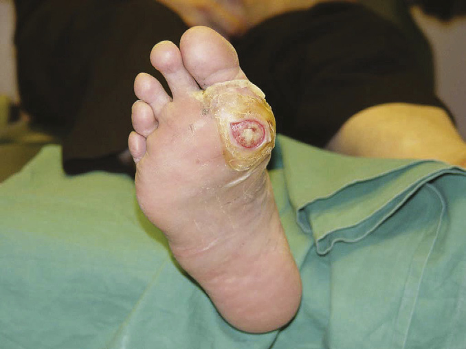
Figure 2 a neuropathic ulcer in a diabetic patient. The breakdown of
skin has occurred at the site of maximum pressure – under the head of
• assessing the ankle jerks.
the first metatarsal joint.
Vascular examination should include palpation for femoral,
popliteal, posterior tibial and dorsalis pedis pulses. Skin colour and temperature, strength of pulsation, and the presence of
Diagnosis and treatment of infection: deep wound swabs often
abdominal and femoral bruits should be documented. Dependent
show the presence of several bacteria, including Gram-positive,
rubor is seen in severe peripheral vascular disease (which may
Gram-negative, aerobic and anaerobic organisms. A positive swab
be mistaken for cellulitis).
may indicate colonization or invasive infection. It is unknown
Clinical assessment should include glycaemic control, dura-
whether superficial ulcers that do not appear infected but which
tion of diabetes, renal disease, cigarette smoking and poor social
have positive swabs should be treated with antibiotics; some
circumstances; these are independent risk factors for complica-
advocate the use of antibiotics more readily in the neuroischaemic
tions of the diabetic foot.
than the neuropathic foot because the former can rapidly lead to necrosis, gangrene and amputation.
Osteomyelitis is a common sequela of diabetic foot ulceration,
Monitoring and self-care are key aspects of management. There
and is usually caused by Staphylococcus aureus. The incidence of
are no data concerning the optimal frequency of specialist review,
methicillin-resistant Staphylococcus aureus in UK hospitals has
although 3–6-monthly review is probably adequate. Patient edu-
more than doubled since 2001.
cation includes washing, inspection, care of corns and calluses,
Plain radiographs should be performed on all patients with dia-
toenail cutting and wearing suitable footwear.
betic foot ulcers, with more sophisticated imaging if osteomyelitis is suspected; MRI and leukocyte scans have equivalent sensitiv-ity (although MRI provides more detailed anatomical information
The ulcerated foot
There may be intrinsic defects in ulcer healing in diabetic patients, including impaired fibroblast function, deficiency in growth factors, and abnormalities of the extracellular matrix. Delayed wound healing and prolonged hospital admissions are there-fore common. A prospective study of 314 consecutive diabetic patients with foot ulcers referred to a multidisciplinary team in a teaching hospital in Sweden reported that healing was achieved in 62% of patients, amputations were necessary in 25%, while 13% died with unhealed ulcers.
Clinical assessment
Neuropathic ulcers are associated with callus, which typi-
cally develops on the plantar aspects of the metatarsal heads
(Neuroischaemic ulcers occur on the margins of the
foot ). Infection is divided into local and superficial;
spreading soft tissue infection and cellulitis; and osteomyelitis.
The signs of inflammation and early infection may be difficult to
detect in the presence of peripheral vascular disease. The abil-
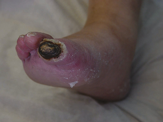
Figure 3 a neuroischaemic ulcer in a diabetic patient. The skin
ity to pass a probe directly onto underlying bone is a sensitive
is necrotic and has developed in an area of relative pressure.
marker of osteomyelitis.
Neuroischaemic ulcers are common on the margins of the foot.
2007 Elsevier ltd. all rights reserved.
and greater specificity). Some groups advocate image-guided
revascularization is planned. Pre-hydration and treatment with
bone biopsy.
N-acetylcysteine is recommended because intravenous contrast
Although surgical intervention is often necessary, conserva-
is nephrotoxic. Magnetic resonance angiography can be superior
tive approaches may be effective. The authors' policy is to use
to conventional angiography and is a useful alternative in renal
MRI to diagnose and assess the extent of osteomyelitis and to
give a 3-month course of appropriate oral antibiotics (initially intravenous only if cellulitis or systemic signs of infection are
Off-loading: the insensate diabetic foot ulcerates as a result
present). MRI is then used to reassess at 3 months:
of minor trauma from poorly fitting shoes and abnormal bio-
• stop antibiotics if the wound has healed and there has been
mechanics. High pressures must be reduced and redistributed in
improvement or resolution of signal change on MRI
order to optimize wound healing.
• repeat the 3-month cycle of antibiotics if the wound is not yet
The first-line treatment in the UK is total-contact casting,
healed and signal change on MRI is improved but persists
which is minimally padded, moulded to the foot, and allows for
• intervention is needed if at any point there is clinical or radio-
mobility while the ulcer heals. Disadvantages include lack of
logical deterioration – revascularization if possible, otherwise
access for wound inspection and dressing, impracticality, and
unsuitability in the presence of ischaemia. Alternative strategies
The choice of antibiotics is governed by the anticipated or proven
include removable cast walkers (e.g. Aircast walker™, Scotch
spectrum of organisms. The regimen of the authors' unit for
cast boots™ (fibreglass removable casts), ‘half-shoes').
emergency management before microbial identification is shown in .
Optimization of wound environment: the goal of preparation
of the wound bed is to create non-infected, well-vascularized
Assessment and treatment of vascular insufficiency: all patients
granulation tissue.
with diabetic ulcers should undergo non-invasive vascular test-
Debridement prepares the wound bed for healing and
ing because clinical evaluation may underestimate the extent of
improves outcome by removing necrotic tissue that often car-
disease. The ratio of blood pressure at the ankle to pressure in
ries the heaviest bacterial load. Surgical debridement is recom-
the arm produces the ankle–brachial pressure index. A value of
mended, although enzymatic debriding agents may also be used.
<0.9 indicates significant arterial disease. Arterial calcification
Sterile larvae of the greenbottle fly (maggots) are effective in
(common in diabetes) may lead to a falsely elevated value. A low
cleaning the wound bed, but may be unacceptable to patients.
value is therefore significant, but normal values can be consis-
Silver- and iodine-containing solutions are used as topical anti-
tent with significant disease.
septics, although efficacy data are lacking.
Waveforms measured by Doppler flow and pulse volume
Other measures – after debridement, tissues should be kept
recording are also helpful. A normal waveform is narrow and tri-
warm and moist to prevent the formation of necrotic material.
phasic: a tall systolic peak, dicrotic notch and rapid run-off. With
Various topical dressings (e.g. alginates, foam, hydrogels, hydro-
increasing disease, the waveform becomes wider and flatter, and
colloids) that absorb wound exudates and promote hydration are
the dicrotic notch is lost, leading to a biphasic (and eventually
available in the UK. Hyperbaric oxygen has been used to coun-
monophasic) pattern. Duplex sonography permits assessment of
teract ischaemia, and there is now evidence for the beneficial use
the anatomy of the vessel and haemodynamic flow. The technique
of vacuum-assisted closure devices.
is operator-dependent at the level of the tibial arteries and below.
Cultured skin dermis (e.g. Dermagraft™) and growth factors
Revascularization with angioplasty or surgery should be con-
may be considered, but are expensive and should be used only
sidered if arterial insufficiency is present. Occlusive vascular dis-
when conventional approaches fail. Cultured skin dermis consists
ease in diabetes involves medium-sized arteries, primarily at the
of neonatal dermal fibroblasts cultured in vitro onto a bioabsorb-
popliteal trifurcation. Distal pedal vessels are often spared occlu-
able mesh to produce metabolically active dermal tissue. Weekly
sive lesions and can be amenable to bypass grafting. Anatomical
application improves wound healing. Synthetic growth factors stim-
evaluation using digital subtraction angiography is necessary if
ulate formation of granulation tissue and enhance epithelialization. Preliminary clinical trials using platelet-derived growth factor and granulocyte colony stimulation factor have been encouraging.
Emergency management of the infected diabetic foot
ulcer before microbial identification
The Charcot foot
Clinical features
The Charcot foot, originally described in association with tabes
First line
augmentin 625 mg tds
dorsalis, affects about 0.2% of patients with diabetes and is
clindamycin 300 mg qds
commonly seen in specialist diabetic foot clinics. Four factors are
ciprofloxacin 500 mg bd
permissive for the onset of Charcot deformity:
Known MRSA carriage
*rifampicin 300 mg bd
• peripheral neuropathy
*Doxycycline 100 mg bd
• autonomic neuropathy • localized osteopenia due to the hyperdynamic flow associated
*assuming previously identified Mrsa was sensitive.
with autonomic neuropathy and resulting in susceptibility to fracture or dislocation
• trauma (significant or minor).
2007 Elsevier ltd. all rights reserved.
Some orthopaedic surgeons suggest doubling the period of
immobilization in diabetic patients with peripheral neuropathy who have a fractured tarsal or metatarsal bone in order to avoid Charcot deformity. The initial insult (which may go unnoticed) results in fracture or dislocation.
The acute phase is characterized by a red, swollen, oedematous
and often painful foot. Foot pulses are bounding. This phase
is often misdiagnosed as cellulitis, deep vein thrombosis, gout
or an ankle sprain. Due to the sensory neuropathy, continued
weight bearing may lead to gross destruction and distortion
of the joint. The mid-foot is usually involved (. The
acute phase may last for months or even years, but eventually
results in increased bone formation, sclerosis and arthrodesis.
In about 20% of cases, the contralateral limb is affected within
several years. The self-limiting nature of the acute phase (and
the association with minor trauma) bears similarities to reflex
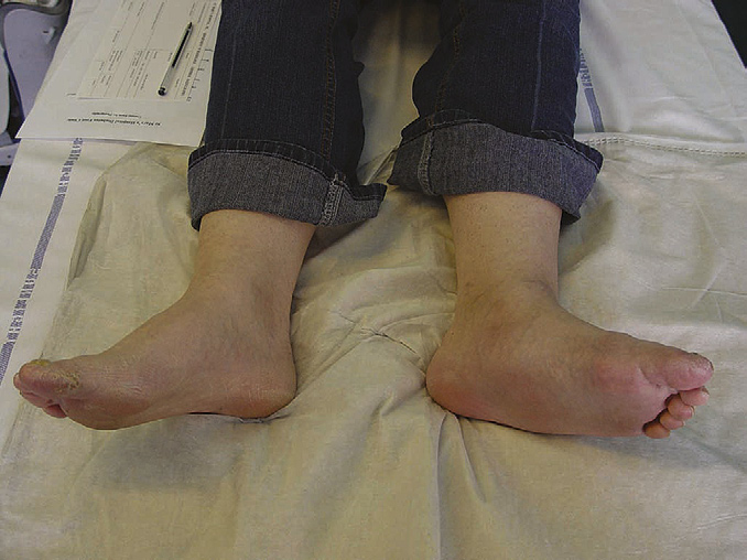
Figure 4 acute charcot changes in the left mid-foot. The mid-foot is
sympathetic dystrophy. Both conditions may respond to bispho-
swollen, hot and deformed. The pathophysiology is complex, but
sphinates given intravenously, possibly through local inhibition
probably involves neuropathy, bone weakening and undetected trauma.
of cytokine release.
The mainstay of treatment in the acute phase is immobiliza-
tion of the joint to prevent further bone destruction and deformity,
MRI. A Charcot foot complicated by ulceration and infection is at
ideally by total contact casting. The optimum duration of immo-
high risk of major amputation.
bilization is not known. The authors have found that a minimum 6-month immobilization is necessary.
Quiescent phase: custom-made footwear can be used. There
Brem H, sheehan P, Boulton aJ. Protocol for treatment of diabetic foot
is a role for reconstructive orthopaedic surgery. Identifying
ulcers. Am J Surg 2004; 187(suppl 1): 1s–10s.
concurrent osteomyelitis (particularly if an area of high pressure
Jeffcoate WJ, Harding KG. Diabetic foot ulcers. Lancet 2003; 361:
has resulted in ulceration) can be particularly difficult, even with
2007 Elsevier ltd. all rights reserved.
Source: http://www.haav24.ee/articles/Seminar/Diabetic_foot_little_review.pdf
expand new drug markets for TB endTB aims to find shorter, less toxic and more effective treatments for multidrug-resistant TB 5 Approach through access to new drugs, a clinical trial, 6 Countries and advocacy at country and global levels. 7 Country requirements Only 11% of multidrug-resistant tuberculosis (MDR-TB) patients get
Frequently asked questions: Deep Brain Stimulation for Parkinson's Disease at UCSF Contents When should one consider surgical therapy? For patients with early Parkinson's disease, levodopa (sinemet) and other antiparkinsonian medications are usually effective for maintaining a good quality of life. As the disorder progresses, however, medications can produce disabling side effects. Many patients on long-term levodopa develop troublesome dyskinesias, excessive movements that often cause the limbs and body to writhe or jump. In addition, their dose


