
Cialis ist bekannt für seine lange Wirkdauer von bis zu 36 Stunden. Dadurch unterscheidet es sich deutlich von Viagra. Viele Schweizer vergleichen daher Preise und schauen nach Angeboten unter dem Begriff cialis generika schweiz, da Generika erschwinglicher sind.
Adipose derived stem and regenerative cells in the treatment of equine orthopedic injuries

Clinical Case Series
Adipose Derived Stem and Regenerative Cells
for the Treatment of
Equine Joint Injuries
Vet-Stem, Inc., 12860 Danielson Court, Suite B, Poway, CA 92064
Developmental bone disease, osteochondritis dissecans (OCD), and subchondral bone
cysts compromise surface cartilage and the underlying supporting bone. Traumatic injuries to joints and their supporting structures happen frequently to high performance athletes as these are the regions under the greatest stress during intense activity. Whether a horse inherits a tendency towards musculoskeletal disease or acquires an injury, if left unchecked, this may lead to degenerative joint disease, osteoarthritis, eventual joint breakdown, and a reduction in functional career longevity.
The application of Stem and Regenerative Cell Therapy in clinical practice has been
utilized in the treatment of tendonitis with the initial source of cells derived from autologous bone marrow. 1 Recently adipose tissue has been described in academic literature as a rich source of Mesenchymal Stem Cells (MSCs).2 Capable of differentiating into multiple cell lines, adipose derived stromal cells (ADAS) have been demonstrated to differentiate into bone, cartilage, tendon, and ligament in both in vitro and in vivo models. 3,4,5
Commercial use of equine adipose derived Vet-Stem Regenerative Cells (VSRC's) has
been available since 2003. Based on patented technology, Vet-Stem, Inc. (Poway, CA) recovers a cell population of multi-potent stem cells, endothelial progenitor cells, pericytes, and associated growth factors from fresh submitted lipectomy samples and returns those cell populations to the practitioner within 48 hours of tissue collection.
The purpose of this series is to review individual clinical case studies and techniques for
utilizing adipose derived stem cells as a therapeutic modality in the treatment of equine orthopedic injuries.

Case 081805-04: 15 Year Old Thoroughbred Broodmare
Bilateral Front Fetlock Osteoarthritis
On August 1, 2005, a 15 year-old Thoroughbred mare presented for bilateral front limb
lameness. Her owners noted that the mare ambulated very little and had progressively lost weight despite the adequate nutrition she had been provided. Previous pain management had included phenylbutazone (2.0 grams BID) followed by naproxen. In addition to causing gastric discomfort, the non-steroidal therapy had proven substandard in managing the mare's discomfort. As she was a valuable broodmare, her owners were concerned about her ability to carry a foal through an entire gestational period.
Physical examination of the mare found her to be moderately underweight, lame at the
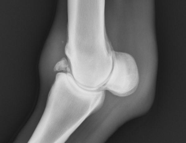
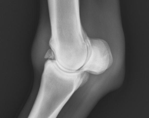
walk (4/5), with effusion of both front fetlock joints. Diagnostic perineural and intra-articular anesthesia isolated the lameness to the fetlock in both front limbs. Radiographic examination revealed chronic fragmentation of the dorso-proximal aspect of the right and left proximal phalanges, along with moderate degenerative joint disease of the right and left metacarpophalangeal joints (Figs 1, 2).
On August 17, 2005, a 16.5 gram sample of subcutaneous adipose was recovered from
the area lateral to the tail head and submitted for stem cell recovery. By 48 hours later, 3.0 million cells were delivered to each of the mare's fetlock joints by intra-articular injection. The patient was maintained on oral non-steroidal anti-inflammatory medications for 48 hours with daily bandage changes. The mare was discharged to her owners with instructions for a return to normal activity and housing.
Figure 1 : Right Front Fetlock
Figure 2 : Left Front Fetlock
Lateral View August 1, 2005
Lateral View, August 1, 2005
Subsequent evaluation at 30 days following stem cell injection found the mare to be
increasingly comfortable, only 2/5 lame at the trot with an estimated weight gain of 150 pounds. No additional pain medications had been administered following the initial 48 hours.

On evaluation 3.5 months following regenerative cell therapy, the attending veterinarian
determined that the mare was sound at the walk and only 1/5 lame in the right front limb at the trot. Despite profound increase in comfort and soundness, evidence of radiographic change was not evident.
Traumatic injuries and degenerative changes to the musculoskeletal system not only
have the potential to end a performance horse's career, they also can limit the equal y important functional capacity of a broodmare's career. Physiologically, pain has been demonstrated to induce elevated circulating levels of Prostaglandin F2α. Elevated levels of PGF2α have been implicated in incomplete corpus luteum function, early embryonic death, and pregnancy loss. The persistent pain of chronic injuries can have a dramatic influence on the reproductive capacity of a broodmare. Adipose derived stem cell therapy has been demonstrated as a valid therapeutic option for the treatment of chronic degenerative joint disease.
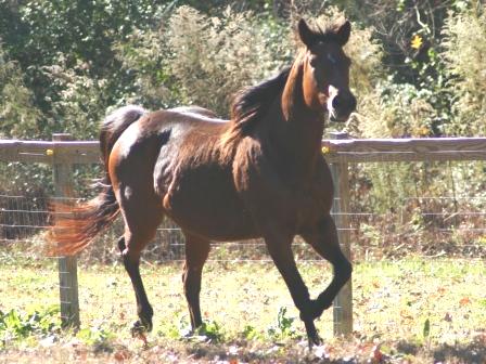
Figure 3 : November 24, 2005
As of November 2005, this mare was sound without the use of non-steroidal anti-
inflammatory medication (Fig 3). She was started under light synchronization protocols in preparation for breeding in 2006.

Case 020805-04: 14 Year Old Mare
Degenerative Joint Disease Secondary to Sepsis
On February 1, 2005, a 14 year-old Thoroughbred mare was presented for evaluation of
a degenerative left tarsal joint due to post injection sepsis. Six months prior to presentation, mepivicaine had been injected into the left tibio-tarsal joint as part of a diagnostic lameness evaluation. Subsequent to injection, the joint became septic and the mare was transferred to a regional referral center. The mare's tibio-tarsal joint was lavaged and treated with a regional anti-microbial infusion for 10 days.
The infectious process eventually subsided with therapy, but the joint developed reactive
proliferation of the articular bony surfaces. The mare presented severely lame on February 1, 2005 for a third and final opinion as the owners were considering euthanasia.
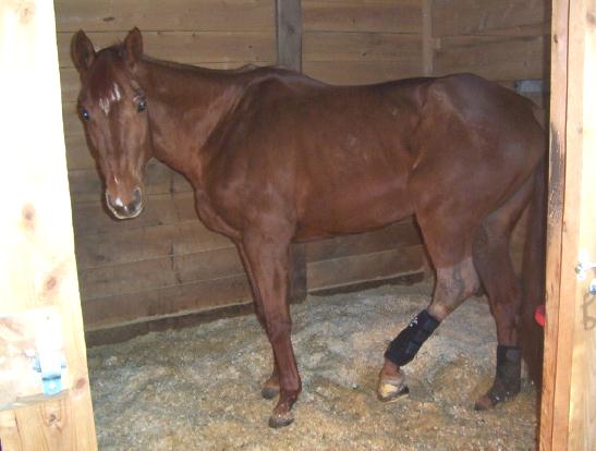
Physical Examination of the mare found her to be non-weight bearing lame (5/5) on the
affected limb (Fig 1). The left tarsal joint had significant joint effusion and the mare was painful
upon manipulation. In addition, the mare was estimated to be 250 lbs. underweight as a result
of the chronic pain. Radiographs of the affected limb revealed diffuse, proliferative osteoarthritis
of the tibio-tarsal joint, with expansion into the proximal and distal intertarsal joints (Fig 2).
Figure 1 : February 2005
Figure 2 : Severe Osteoarthritis
Left Tarsus, February 2005
Figure 3: May 2005
On February 7, 2005 twenty-two grams of subcutaneous adipose tissue was excised
from over the right hip and submitted for stem cell recovery.
On February 9, 2005 10 million adipose derived stem cells were delivered to the tibio-
tarsal joint by intra-articular injection using aseptic technique. The mare was continued on her daily anti-inflammatory regimen of up to 4 grams of phenylbutazone to decrease the overall chronic pain. Approximately 10 days post treatment she began showing signs of increased comfort and ambulation. A second stem cell preparation of previously frozen cells containing 4.5 million cells was administered on April 1, 2005 and the mare continued to show increasing comfort, as well as decreased effusion of the affected joint. As her condition continued to improve, her daily NSAID requirements continued to decrease.
By May 2005, the mare was ambulating comfortably, was being turned out daily, and
had gained significant weight since the original evaluation (Fig 3). Her comfort level had increased so dramatically that her only anti-inflammatory requirements were a tapered dose of meclofenamic acid every 3 to 4 days as needed.
This mare continued to improve to the point that in July, 2005 she was returned to light
riding, an activity not performed since 2004 (Fig 4).
Figure 4: July 2005
Case 113004-02: 2 Year Old, Arab Gelding
Sub-Chondral Bone Cyst Causing Lameness
On September 15, 2004, a 2 year-old Arabian gelding, was presented for a lameness of
the right hind limb. Physical examination and regional anesthesia isolated the discomfort distal
to the right hind fetlock joint and digital radiographic evaluation revealed a sagittal, proximal
sub-chondral bone cyst (OCD lesion) of the second phalanx. The patient was discharged with
instructions for stall rest and a re-evaluation in 60 days was scheduled. Follow up evaluation
on November 24, 2004 revealed that the lameness had continued to worsen and that the sub-
chondral bone cyst had increased in size (Fig 1). As the sub-chondral bone cyst was unlikely to
spontaneously resolve, the owners elected to treat the affected joint with adipose derived
regenerative cell therapy.
On November 29, 2004, the gelding was sedated and a local anesthetic was
administered over the right hip, lateral to the tail-head. Fifteen grams of subcutaneous fat was removed by lipectomy, packaged, and sent via overnight courier for regenerative cell recovery. On December 1, 2004 the horse was anesthetized using a modified injectable induction protocol and maintained on isflourane and oxygen. The right rear limb was prepared using sterile technique and the affected region was desensitized using regional nerve anesthesia. Peri-articular access to the subchondral bone cyst was created using an orthopedic drill via a dorsal approach where 4.8 million cells were delivered utilizing bone marrow as a carrier agent. The delivery port was closed and the surgical site was closed using surgical skin staples.
Radiographic evaluation 36 days following regenerative cell therapy revealed significant
improvement and greater than 50% increase in radio-opacity of the sub-chondral defect (Fig 2).
Figure 1 :
Figure 2 :
Figure 3 :
roximal Sub-Chondral Bone
Proximal Sub-Chondral Bone
Proximal Sub-Chondral Bone
st of the Second Phalanx
Cyst of the Second Phalanx
Cyst of the Second Phalanx
November 2004
January 2005
July 2005
The horse continued recommended physical therapy and returned to competitive activity
within 45 days of stem cell therapy. Radiographic evaluation of the right hind limb in July 2005 revealed resolution of the subchondral defect. The underlying cancellous bone showed adequate opacity and the chondral surfaces showed no indication of compromise (Fig 3). At the time of this evaluation, the colt had returned to full activity and was actively competing.
Case 070104-01: 18 Month Old Reining Horse
Intra-Lesion Injection of Sub-Chondral Bone Cyst
Concurrent with Arthroscopic Surgery
On June 30, 2004, an 18 month-old Quarter Horse presented for pre-training screening
films. This decision was made based on the gelding's familial history: Two full brothers both had subchondral bone cysts of the stifle joints. Lameness examination found this horse to be sound at a walk, trot, and following upper limb flexion. Despite a normal physical exam, radiographs of the right hind stifle joint revealed abnormal flattening of the cartilage surface and the presence of a moderate sub-chondral bone cyst within the medial femoral condyle (Fig 1). Because of familial history and radiographic findings, surgery and concurrent direct intra-cystic injection of adipose derived regenerative cells was the elected therapy. On June 30, 2004, 17.6 grams of subcutaneous adipose tissue was harvested from the gluteal region lateral to the tail head and submitted for stem cell recovery.
Figure 1 : Radiograph Right Stifle Figure 2 : Radiograph Right Stifle
June 30, 2004 December 16, 2004
On July 2, 2004 exploratory arthroscopic surgery of the right medial femoro-patellar joint
identified moderate damage to the surface cartilage. After debridement of necrotic tissue, the joint was lavaged and then decompressed. Immediately prior to scope removal 7.1 million adipose-derived regenerative cells were injected into the cystic remnant and the surgical ports were closed by standard technique. The patient was discharged with instructions to restrict activity to stall rest only for the initial 30 days following surgery. This was to be followed by 5-10 minute hand walking sessions for 30 days and then gradual increases in exercise were permitted over the next 4 months.
Physical examination on December 16, 2004 found the horse to be sound at the walk,
trot, and following upper limb flexion. Radiographic evaluation revealed a 50% reduction in size of the sub-chondral bone cyst (Fig 2).
Figure 3: Radiograph Right Stifle
March, 2005
Based on clinical improvement, the owners elected to pursue a second arthroscopic
guided regenerative cell injection of the affected joint. Adipose tissue was recovered in the identical prior fashion as on July 2004 and 5.5 million cells were returned for surgical delivery on December 22. Arthroscopic evaluation of the medial femoro-patellar joint revealed greatly improved visual characteristics of the cartilage surface where the previous lesion was located. As gross compromise to the cartilage surface could not be visualized, the second application of adipose derived stem cells was administered via intra-articular deposition only. The patient was discharged with instructions to gradually resume the original rehabilitation program.
Over the next 3 months, the patient was exercised daily. This gelding progressed
quickly from rehabilitation status into a conditioning program and formally started training in March 2005. Radiographic evaluation revealed upwards of 75% resolution of the subchondral defect (Fig 3) and physical examination revealed the horse to be sound. The horse has maintained soundness for over 8 months.
Case 020105-02: 2 Year Old Cutting Horse
Bilateral Sub-Chondral Bone Cysts
Concurrent with Arthroscopic Surgery
On January 31, 2005, a 2 year-old Quarter Horse mare presented for pre-training
screening films. Lameness examination found this horse to be sound at a walk, trot, and following upper limb flexion. Despite a normal physical exam, radiographs of both stifle joints identified abnormal flattening of the cartilage surface and the presence of bilateral sub-chondral bone cysts within both medial femoral condyles (Fig 1).
Figure 1 : Radiograph Right Stifle
January 31, 2005
Based on radiographic findings, surgery in conjunction with direct intra-cystic injection of
adipose derived stem cells was elected by the owners and attending veterinarian. On January 31, 2005, 29.5 grams of subcutaneous adipose tissue was harvested from the left gluteal region lateral to the tail head and submitted for stem cell recovery.
On February 2, 2005 exploratory arthroscopic surgery identified surface cartilage lesions
in both medial femoro-patellar joints with the right more severely affected than the left (Fig 2, 3). Necrotic tissue was debrided, both joints were lavaged, and then decompressed.
Figure 2: Feb 2, 2005
Figure 3: Feb 2, 2005
Arthroscopic Image Right MFT Joint
Arthroscopic Image Left MFT Joint
Immediately prior to scope removal 5.5 million adipose-derived regenerative cells were
injected into each cystic remnant and the surgical ports were closed by standard technique. The patient was discharged with instructions to restrict activity to stall rest only for the initial 30 days following surgery, and then to permit 5-10 minute hand walking sessions for 30 days.
Over the following 3 months, the patient was exercised daily and progressed from
rehabilitation status into a reconditioning program; this cutting horse formally entered training in
May 2005. Radiographic evaluation on May 3, 2005 revealed upwards of 80% resolution of the
subchondral defect in the right stifle and 70% resolution in the left (Fig 4). Physical
examination revealed the horse to be sound at the walk, trot, and following upper limb flexion.
By August 2005, the horse had been actively training for 3 months. To date, this horse has
shown no lameness.
Figure 4 : Radiograph Right Stifle
May 3, 2005
Case 010505-02: One Year Old Warmblood Filly
Shoulder Osteochondrosis Dissecans
On October 22, 2004, a one-year-old Warmblood filly was presented with a grade 2/5 left
front lameness. The injury occurred two weeks prior to presentation during a training session. The attending clinician performed regional nerve and intra-articular blocks with carbocaine to isolate the lameness to the shoulder. Ultrasound revealed mild roughening of the median tubercle of the humeral tuberosity and significantly increased amount of fluid in the bicipital bursa of the left shoulder. On radiographs there were no bony changes noted. At this time, the decision was made to inject the bursa with 2 mls of Hylartin-V®, 5 mg of Triamcinolone Acetonide and 60 mg of Depo-medrol; in addition, stall rest was prescribed.
On December 30, 2004, the Warmblood filly was reevaluated for a persistence of the
same lameness. The attending veterinarian determined that arthroscopy combined with stem cell therapy was the best choice at this time to facilitate healing. On January 4, 2005, 13.68 grams of subcutaneous adipose tissue was collected from just lateral to the tail head and submitted for recovery of regenerative cells. On January 7, 2005, arthroscopy was performed to evaluate the joint and to debride the lateral glenoid and humeral head OCD lesions. The OCD lesions involved approximately 1/3 of the mid-lateral aspects of the humeral head and the glenoid; they did not involve the major weight bearing surface of either structure and the remainder of the joint appeared in excellent condition. After debridement and lavage, 2 mls of regenerative cells (8.7 million total cell count) were instilled into the joint at the site of debridement. The prognosis for returning to prior level of performance was guarded to good with a 65% chance of long term athletic soundness.
On July, 2005, a six month post-treatment examination was performed. Left shoulder
radiographs showed a flattening of the previous OCD area of the humeral head. She was still slightly lame (1/5 on left shoulder during work outs) and was started on a series of chondroprotective agents (Legend® and Adequan®).
One year post-injury, the filly was presented for follow-up physical examination in
addition to arthroscopy and joint evaluation. She was sound on the left front and arthroscopy revealed smooth cartilage covering all of the humeral head and glenoid fossa OCD debridement areas. No further joint debridement was determined necessary, the joint was closed and the patient was maintained on oral non-steroidal therapy for three days post exam.
In June 2006, the Warmblood fil y's owner reported that they had her under saddle
during the prior 30 days and that she had been sound and performing well. She was awarded the "Best Young Horse" at each of her last 3 In-Hand competitions. She was entered into the International Hunter Futurity Two-Year-Old Under Saddle class in August.
Case 062905-01: Two Year Old Quarter Horse Colt
Stifle Osteochondrosis Dissecans
On June 21, 2005, a two-year old Colt was presented with a grade 2/5 left hind
lameness. On high flexion test the left hind became a 3.5/5 lame. The attending clinician performed regional nerve and intra-articular blocks with carbocaine and isolated the lameness to the left stifle. Further diagnostic evaluation with digital radiographs showed mild to moderated flattening of the medial femoral condyle. The attending veterinarian determined that arthroscopy combined with regenerative cell therapy was the best choice at this time to facilitate healing.
On June 28, 2005, 16.87 grams of subcutaneous adipose tissue was collected from just
lateral to the tail head and submitted for recovery of regenerative cells. On July 1, 2005, arthroscopy was performed on both stifle joints. The left stifle required debridement of a chondromalasia lesion of the medial femoral condyle. This lesion was moderate-sized and was in close proximity to the major weight-bearing surface of the medial aspect of the joint. There were no chondromalasia lesions of the lateral femoral condyle noted. Micro fracture of the subchondral bone of the medial femoral condyle was performed. The right stifle chondromalasia lesion of the medial femoral condyle was approximately 10-15% smaller than that found in the left. Microfracture of the subchondral bone was performed. Adipose derived regenerative cells (4.3 million each site) were injected into the medial femorotibial joint prior to closing. Prognosis for a long term athletic soundness was said to be 80%
On February 2, 2006 a reevaluation was performed. Prior to this exam two series of
Legend® and Adequan® injections had been administered. The patient was sound when trotted in hand and 1.5/5 lame on upper limb flexion test. On August 9, 2006 the colt was observed to be sound in both hind limbs and on all components of the physical exam.
On August 23, 2006, the horse had continued to be sound for six months and a one-year
follow up reevaluation with arthroscopy was performed. The left medial femoral condyle cartilage repair tissue appeared thin and somewhat translucent. The defect was completely covered; however, some cartilage fracture lines were still present in the debrided area of the remaining cartilage. There was minimal synovial inflammation noted in the left stifle. The right medial femoral condyle cartilage repair tissue appeared thicker than on initial arthroscopy and was nearly flush to the surrounding cartilage. The defect was completely covered with repair cartilage that was relatively smooth with a swirling fiber appearance. As with the left stifle, the right stifle showed minimal synovial inflammation
Presently, the colt is in training and has not presented any lameness issues.
Note: Images not available due to computer hard-drive failure at clinic.
Western Pleasure Quarter Horse
MTIII Distal Fracture Involving the Joint
A Western Pleasure Quarter Horse was presented for acute lameness during workouts
on December 13, 2005. The patient was a grade 3/5 lame on the left hind limb. Based on pain elicited in addition to visible and palpable swelling, the attending veterinarian chose to radiograph the left hind fetlock. On radiographic review, a fracture to the medial condyle in MTIII at the joint surface was noted.
The fetlock was injected with a combination of Hylartin-V® and amikacin and two
months stall rest was prescribed. On reexamination February 6, 2006, the Quarter Horse was still grade 3/5 lame on the left hind. Radiographs taken on this day showed the continued presence of the fracture and when the fetlock was blocked with carbocaine, the horse went sound. At this time, the veterinarian and owner elected to administer regenerative cell therapy.
On February 7, 2006 a 19.76 gram sample of subcutaneous adipose tissue was
harvested from the tailhead region and submitted for recovery of regenerative cells. On
February 9, 2006, 7.4 million regenerative cells were sterilely injected into the left hind fetlock
joint.
December 13, 2005 April 4, 2006
pril 4, 2006
On March 7, 2006, the horse was reevaluated thirty days post-injection of stem cell
therapy. No lameness was noted with hand trotting and the patient was 2/5 lame on flexion.
Aquatred rehabilitation was prescribed at this time.
On recheck exam April 4, 2006, the patient continued to show no lameness at the trot and was
minimally (1/5) lame on flexion. The patients rehab schedule was expanded to include ground
work. On June 1, 2006 a final recheck was performed; the patient was sound on flexion as well
as when trotted in hand. No other treatments were recommended.
This Quarter Horse is currently being ridden Western Pleasure and remains sound with
no lameness or pain noted by owner or veterinarian.
Case 092209-02: Five year old Quarter Horse Mare
Bilateral Stifle Osteoarthritis
A five year old Quarter Horse mare was presented with a chronic (8 month) history of
hind limb lameness. The patient would block out sound when the stifles were injected with carbocaine. Prior therapies had included (individually or in combination): Hylartin-V®, adequan®, amikacin, and shockwave. Severe joint effusion had been noted after each treatment and on fluoroscopy no bony changes were visible in the joint. With consideration of the chronicity of the joint in combination with the owners declining to continue trying further conventional therapies such as steroid injections or surgery, the attending veterinarian elected to use regenerative cell therapy.
On September 21, 2005 a 12.91 gram sample of subcutaneous fat was removed from
the lateral side of the mare's tail head and was sent for collection of regenerative cells. On September 23, 2006, one million cells were injected via sterile preparation into each stifle joint. Stall rest was prescribed and a reevaluation scheduled for two months later.
On November 23, 2005 no lameness was noted when longed and no stife joint effusion
was palpated. Due to marked improvement, the rehabilitation program was expanded to include legging up.
On February 14, 2006, a final set of radiographs showed no progression of osteoarthritis
and the Quarter Horse mare returned to full activity. Since that time, the mare has remained sound and the owner reports that she has regained her prior level of performance in cutting work.
Note: Images not available due to computer hard-drive failure at clinic.
Case 111005-04: A Four Year Old Quarter Horse Gelding
Bilateral Osteoarthritis of Front Coffin Joints
On November 2, 2005 a four-year-old Quarter horse gelding presented with a one year
history of front limb lameness that had been treated multiple times with steroid injections. The patient was a grade 3/5 lame in the left front; when the left front coffin joint was blocked with carbocaine, the patient went sound at the trot. A series of x-rays were taken which showed changes around the navicular bursa and the distal interphalangeal joint of the left front limb. Severe osteoarthritis was diagnosed and the left front coffin joint was treated with Hylartin. Concerned for the chronicity of the lameness and hoping to regain more long term function for the patient, the attending veterinarian also chose to administer regenerative cell therapy.
On November 9, 2005 a 17.66 grams of subcutaneous adipose tissue was obtained
lateral to the gelding's tail head and was sent for collection of regenerative cells. On November 11, 2005 each front coffin joint was injected sterilely with 2.3 million regenerative cells (the right joint was injected as a prophylactic measure). The patient was prescribed stall rest in conjunction with a light turnout for the following two months.
On reevaluation January 5, 2006, the veterinarian detected no lameness at either the
walk or trot; flexion tests were negative for both front limbs. At this time light riding was added to the rehabilitation schedule. On January 13, 2006, no lameness was noted in either limb and both joints received a routine injection of Hylartin. On February 8, 2006 the patient continued to exhibit no sign of lameness on a lunge line and was negative with hoof testers. At that time, the gelding was in a full training regimen for reining and had not taken another lame step.
During an April 4, 2006 conversation, the owner told the veterinarian that the gelding had
been very sound and had competed three times with wonderful results. He does not seem to be sore at all, is feeling great and is a pleasure to show. Prior to the stem cell injection, every time we tried to step him up to get ready to show, he would go lame. Now, since the stem cell therapy, he has not had any lameness problems".
Case 122005-03: Two Year Reining Quarter Horse Colt
Bilateral Subchondral Bone Cysts
A two-year-old Quarter Horse was presented for lameness evaluation on April 15, 2005.
On examination, the veterinarian noted that the patient stepped short on the left rear and was positive to high flexion test of same limb. A mild amount of joint effusion was palpated over the left femoral patellar joint. Fluoroscopy was performed on both stifles. Sclerosis and a cyst of the right medial femoral condyle were also noted. No treatments were administered at this time. The colt was prescribed calcium carbonate and a 30 day recheck was recommended.
Six months later, on December 15, 2005, a reevaluation was done: the patient was
short on the right hind and positive to flexion test on both stifles. In order to more fully diagnose the etiology of the lameness of this Quarter horse, fluoroscopy of both stifles was performed and revealed sclerosis of both medial femoral condyles. Both stifles were injected with Hylartin and amikacin. At this time the veterinarian and the colt's owners elected to use regenerative cell therapy to treat the stifle joints.
On December 20, 2005 a 22.49 gram sample of subcutaneous adipose tissue was taken
and sent for collection of regenerative cells. On December 22, 2005 3.1 million regenerative cells were sterilely injected into each stifle joint. Post-injection, the patient was prescribed stall rest for 60 days.
On February 16, 2006, no lameness was detected while the colt was on longe line; he
was also negative to flexion tests.
On reevaluation May 23, 2006, the colt still exhibited no lameness. Digital radiographs at this time showed marked density of the left rear medial femoral cyst. The right stifle also showed improvement as the density of the area of the medial femoral condyle had improved significantly on follow-up radiographs compared to the initial images.
Pre-Treatment, December 15, 2005
5 Months Post Treatment May 23, 2006
The Quarter Horse colt returned to full training and was entered in a futurity in Oklahoma city in November 2006.
References: 1 Herthel DJ. Enhanced suspensory ligament healing in 100 horses by stem cells and other bone marrow components. Proceedings, American Association of Equine Practitioners, 2001; 47:319-321.
2 Zuk PA, Zhu M, Ashjian P, et al. Human adipose tissue is a source of multipotent stem cells. Molecular Biology of the Cell 2002; 13: 4279.
3 Hicok KC, Zhou Y, Pucilowski Y. Adipose derived adult stem (ADAS) cells differentiate into bonesynthesizing osteoblasts in vivo. Presentation #M233
4 Nathan S, Das De S, Thambyah A, et al. Cell-based therapy in the repair of osteochondral
defects: a novel use for adipose tissue. Tissue Eng. 2003 Aug;9(4):733-44.
5 Quintavalla J, Uziel-Fusi S, Yin J, Boehnlein E, et al. Fluorescently labeled mesenchymal stem
cells (MSCs) maintain multilineage potential and can be detected following implantation into
articular cartilage defects. Biomaterials. 2002 Jan;23(1):109-19.
**Vet-Stem would like to thank the clinicians and support staff of the following clinics for their
contribution of case data and images: Cave Creek Equine Surgical Center, Performance
Equine Associates, Weatherford Equine Medical Center, Brazos Valley Equine Hospital, Great
Basin Equine, Pioneer Equine Clinic, Dr. Van Snow, Equine medical Associates.
Source: http://www.australianstemcells.com.au/attachments/article/202/Equine%20Joint%20Injuries%20-%20Clinical%20Case%20Series.pdf
Cold Fusion Cloning Kit Cat. #s MC010A-1, MC100A-1, MC101A-1 User Manual Store the master mixture and positive controls at -20˚C Store the competent cells at -80˚C. A limited-use label license covers this product. By use of this product, you accept the terms and conditions outlined
HSI HEALTH SCIENCES INSTITUTE MEMBERS ALERT FOR NOVEMBER 2004 HSI Advisory Panel One-time charge for long-time relief Medical Adviser, Martin Milner, N.D.Professor, National College of Naturopathic from chronic pain Medicine; President, Center for Natural Medi-cine, Portland, OR; www.cnm-inc.com


